Cellular Thermoprobe for Fluorescence Lifetime, 200 ug
Cellular Thermoprobe for Fluorescence Lifetime (FLIM-Type)
Cat No. FDV-0004_200 ug, FDV-0004_200 ug x3
Size 200 ug, 200 ug x3
Storage RT, Dark Storage
| Product No. | Size | Link |
| FNK-FDV-0004-200 | 200 ug |  |
| FNK-FDV-0004-200x3 | 200 ug x3 | |
Description
- Catalog Number: FDV-0004
- Lot Number: see vial label
- Size: 200ug or 200 ug x3
- Average Molecular Weight: 12,300
- Purity: >99%
- Appearance: Yellow powder Solubility: Soluble in water
- Spatial Resolution: 200 nm
- Temperature Resolution: 0.05-0.54℃
- License: This product is licensed by Tokyo University and KIRIN Co., Ltd.
Product Background
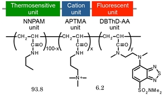
“Cellular Thermoprobe for Fluorescence Lifetime” is a fluorescent polymeric thermometer for living cells. It diffuses throughout the cells and gives the information about intracellular temperature distribution by fluorescence lifetime imaging microscopy. “Cellular Thermoprobe for Fluorescence Lifetime” can be delivered into cell without microinjection. It is highly photostable and easy-to-use. With its cell permeability, “Cellular Thermoprobe for Fluorescence Lifetime” is applicable for both adherent and suspension cells. It enables us to distinguish intracellular temperature in cultured mammalian cells at organelle level. For instance, the previous report demonstrated that the average temperature difference between the nucleus and the cytoplasm was 0.98 ℃ (reference 1). “Cellular Thermoprobe for Fluorescence Lifetime” is an innovative new reagent that provides unprecedented scientific insight.
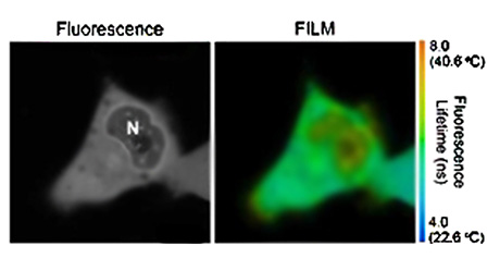
Temperature mapping in living HeLa cells.
Confocal fluorescence image (left) and fluorescence lifetime images (right) of the “Cellular Thermoprobe for Fluorescence Lifetime” in HeLa cells. N indicates the nucleus.
Storage
- Shipping: Shipped at ambient temperature
- Storage: Store at ambient temperature (powder). For reconstituted solution, store at +4℃. Protected from light.
- Before open the top, spin the vial down briefly.
- Reconstitute 200?g powder of “Cellular Thermoprobe for Fluorescence Lifetime” in 20?l of ultrapure water*1.
- Dissolve it completely by vortex or tapping.
- Store the stock solution (1% w/v) at +4℃. Protect from light. Note that the stock solution needs to be incubated at +4℃ at least overnight before proceeding to experiment to obtain full extension of the polymer.
*1 Ionic solutions such as DMEM and PBS inhibit the incorporation of Cellular Thermoprobe for Fluorescence Lifetime. If you find poor solubility, put it on ice for a while until it dissolve.
Representative absorption and fluorescence spectra of DBThD-IA.
The absorption spectra were measured in acetonitrile (black). The fluorescence spectra were measured with excitation at 458 nm in ethyl acetate (red), acetonitrile (orange) and methanol (blue).
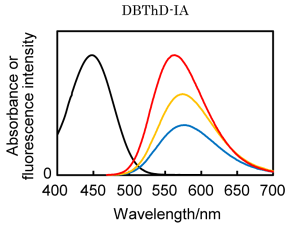
Optimal excitation and emission will be determined by your own. As an example, excitation at 405 nm and emission at 560-610 nm would work well.
Preparation of cell extract for calibration curve
- Cell pellets (1 x 10⁷) were collected from 100 mm dish and resuspended in hypertonic buffer (2.5 ml, containing 0.42 M KCl, 50 mM HEPES-KOH, 5 mM MgCl2, 0.1 mM EDTA, 20% glycerol, pH 7.8).
- Lyse cells using a 25-G needle with a syringe.
- Centrifuge (11,000 r.p.m., 15 min, 4℃) and collect the supernatant.
- Dilute the supernatant with water up to 40% to adjust its KCl concentration to 0.15M.
- Dilute 1ul of “Cellular Thermoprobe for Fluorescence Lifetime” in water (1% w/v) with cell extract (20-100 u).
- Put the solution on a glass bottom dish.
- Set the temperature of the stage heater at the lowest you can (e.g. 25℃).
- Measure the fluorescence lifetime after the medium temperature becomes steady.
- Adjust the medium temperature at your choice (e.g. 26℃).
- Measure the fluorescence lifetime after the medium temperature becomes steady.
- Repeat step 5-6 until reaching the maximum temperature of the stage heater.
- Plot the fluorescence lifetime against temperature to obtain a calibration curve. Estimate the temperature of your sample based on the calibration curve.
*1 Calibration curve can be also generated by Spectrofluorometer or Fluorescence Plate Reader equipped with temperature control.
Introduction of “Cellular Thermoprobe for Fluorescence Lifetime” into Suspension Cells
- Collect the suspension cells by centrifugation at 400 x g for 3 min and wash it with 1 ml of a 5 % glucose solution and centrifuge it.
- Remove the supernatant.
- Resuspend the cell pellets in a 5 % glucose solution at a density of 1 x 10⁶ cells/ml.
- Add “Cellular Thermoprobe for Fluorescence Lifetime” in water (1% w/v) to a 20-100 fold*1 volume of cell suspension.
- Incubate the cells at 25℃ for 10 min.
- Centrifuge it and remove supernatant, and add 1ml PBS.
- Centrifuge it and remove supernatant, and resuspend in PBS.
- For the fluorescence imaging, approximately 10?l of the cell suspension is dropped onto a coverslip and observe it immediately*2.
*1 Optimal dilution rate of “Cellular Thermoprobe for Fluorescence Lifetime” depends on cell types.
*2 Set the approrpriate temperature (e.g. 32 ~ 33℃) in a microscope cage incubation chamber based on your calibration curve and/or experimental condition.
Introduction of “Cellular Thermoprobe for Fluorescence Lifetime” into Adherent Cells
- Prepare the cells at the 30 to 50 % confluency on glass bottom dish or equivalent.
- Remove the medium and wash with a 5 % glucose solution*1.
- Add 0.01-0.05 w/v%*2 of “Cellular Thermoprobe for Fluorescence Lifetime” in 5 % glucose solution*1*3.
- Incubate the cells at 25℃ for 10 min.
- Wash the cells with PBS three times.
- Add phenol red-free culture medium and measure the fluorescence lifetime with appropriate temperature in a microscope cage incubation chamber*4.
*1 In the case that the dissociation of the adherent cells were observed in 5% glucose solution, 5% glucose solution with 0.1 – 0.3 mM CaCl2 may improve it.
*2 Optimal dilution rate of “Cellular Thermoprobe for Fluorescence Lifetime” depends on cell types.
*3 The volume of the solution depends on the culture dish type. But we assume that minimum amounts of volume (50-100 ul) is sufficient to measure the cellular temperature.
*4 Set the appropriate temperature (e.g. 32 ~ 33℃) in a microscope cage incubation chamber based on your calibration curve and/or experimental condition.
Note
Above methods (Reconstitution, Preparation of cell extract for calibration curve, How to generate calibration curve and introduction) should be optimized depending on the cell type and organisms you use.
Example of Calibration Curve in HeLa cell extracts
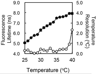
Fluorescence response (closed, left axis) and temperature resolution (open, right axis) in HeLa cell extracts. The temperature resolution of Cellular Thermoprobe for Fluorescence Lifetime was 0.05 – 0.54℃ in the temperature range between 28 and 38℃.
Reference
- Hayashi T, et. Al., PLoS ONE. 2015 Feb 18; 10(2): e0117677. doi:10.1371/journal.pone.0117677
- Kimura H, et al., Sci Rep, . 2017 7(1):12978.
- Hoshi Y, et al., J Neurosci. 2018 38(25):5700-5709.
- Halip H, et al., Methods Appl Fluoresc. 2020 8(2):025003.
- Kato H, et al., Life Sci Alliance 2020 3(3):e201900576.