


Human Lumican Antibody
Human Lumican Antibody
General information
| Cat. No. | :FNK-FDV-0001 |
| Size | :25ul |
| Antigen Species | :Human |
| Host Species | :Rabbit |
| Cross Reactivity | :Human,Rat |
| Purification | :Affinity Purified |
| Immunogen | :Synthetic Peptide (aa211-227 of Human Lumican) |
| Application | :IHC-P: 1:200 to 1:2,000, Western Blotting: 1:1,000 |
| Formulation | :PBS (10mM Phosphate buffer, 0.15M NaCl (pH7.2))(0.3mg/ml) |
| Preservative | :0.1% Sodium Azide |
| Storage | :-20ºC, Avoid freeze/thaw cycles. |
Description
Lumican is a glycoprotein of 338 amino acids, which is a member of small leucine-rich proteoglycan (SLRP) family. This glycoprotein has small core proteins of approximately 40 kD and the core protein contains a central region of leucine-rich repeats flanked on either side by a disulfide-bonded domain. The central region of the molecule possesses four asparagine residues capable of participating in N-linked glycosylation, which regulates the assembly and diameter of collagen fibers. Lumican has an important role for such as cell proliferation, cell migration and cell adhesion. Recently, lumican overexpression has been reported in various cancer tissues, such as carcinoid tumor, breast, colorectal, neuroendocrine cell, uterine cervical, lung and pancreatic cancers.
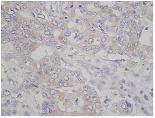
Human pancreatic cancer (formalin-fixed, paraffin embedded) stained with human lumican antibody (#FDV-0001).
Applications
|
Immunohistochemistry
|
|
 |
|
|
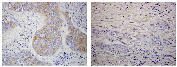
Human lung cancer (formalin-fixed, paraffin embedded) stained with human lumican antibody (#FDV-0001). Lumican is observed in cancer cells (left image), and interstitium (right image) in lung squamous cell carcinoma
|
|
|
Western Blotting
|
|
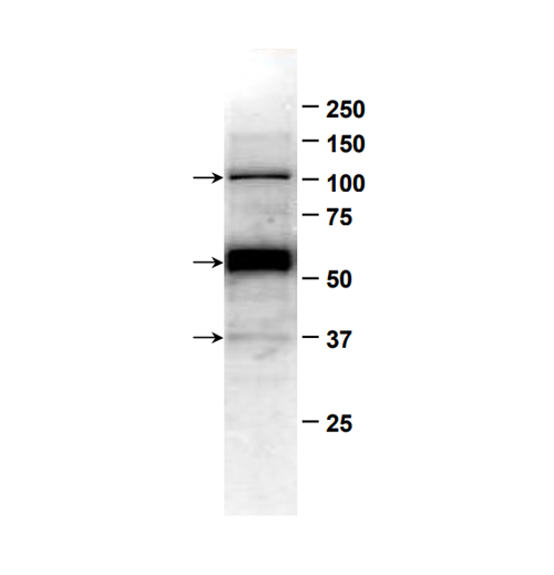 |
Western blot analysis on A549 human lung cancer cell lysate using human lumican antibody (#FDV-0001). 37KDa band of lumican core protein and glycosylated forms of lumican are observed. |
References
1. Yamamoto T, et al., Cancer Lett. (2012) 320, 31-39, [PMID: 22266188]
2. Ishiwata T, et al., Exp Mol Pathol. (2010) 88, 363-370.[PMID: 20138170]
3. Matsuda Y, et al., Int J Oncol. (2008) 33, 1177-1185[PMID: 19020750]
4. Ishiwata T, et al., Oncol Rep. (2007) 18, 537-543[PMID: 17671699]
5. Seya T, et al., Oncol Rep. (2006) 16, 1225-1230 [PMID: 17089042]
6. Shinji S, et al., Int J Oncol. (2005) 26, 873-880 [PMID: 15753980]
7. Kusafuka K, et al., J Pathol. (2004) 203, 953-960[PMID: 15258999]
8. Ishiwata T, et al., Pathol Int. (2004) 54, 77-81[PMID: 14720136]
9. Saika S, et al., Invest Ophthalmol Vis Sci. (2003) 44,2094-2102 [PMID:12714648]
10. Naito Z, et al., Exp Mol Pathol. (2003) 74, 33-39[PMID: 12645630]
11. Lu YP, et al., Pathol Int. (2002) 52, 519-526[PMID: 12366811]
12. Naito Z, et al., Int J Oncol. (2002) 20, 943-948[PMID: 11956587]
13. Onda M et al., Exp Mol Pathol. (2002) 72, 142-149[PMID: 11890723]
14. Ping Lu Y, et al., J Pathol. (2002) 196, 324-330[PMID: 11857496]
15. Qin H, et al., J Pathol. (2001) 195, 604-608[PMID: 11745697]
16. Baba H, et al., Jpn Circ J. (2001) 65, 445-450[PMID: 11348051]
Aliases for LUM Gene
