

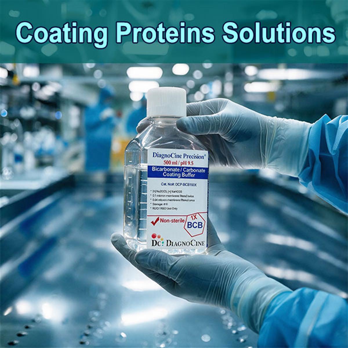
Bicarbonate / Carbonate Coating Buffer — A Guide for the Research Laboratory
One formulation, ready to use at pH 9.6. Go straight to the product page, or read on for the adsorption chemistry, the full application list, and the coating conditions that decide whether an assay works.
| Coating Buffer | Composition · pH | Primary Use | Product Page |
|---|---|---|---|
| Bicarbonate / Carbonate Coating Buffer [1X] | Sodium carbonate / sodium bicarbonate · pH 9.6 ± 0.1 | Passive immobilization of antibody or antigen on polystyrene for solid-phase immunoassay | Viewarrow_forward |
Looking for a specific format? See the full application list · Browse applications by format family · Read the FAQ
The step that sets the ceiling on the whole assay
A carbonate-bicarbonate coating buffer is an alkaline solution, classically 0.05 M sodium carbonate and sodium bicarbonate at pH 9.6, used to immobilize a capture antibody or antigen onto a hydrophobic plastic surface by passive adsorption. It is the first step of most solid-phase immunoassays and sets the ceiling on everything downstream: uneven or inefficient coating cannot be recovered by later optimization. The same alkaline chemistry also serves a second role in the lab as the reaction environment for amine-reactive labeling. Both uses are covered below.
- One formulation stocked: carbonate-bicarbonate coating buffer, 1X — sodium carbonate / sodium bicarbonate at pH 9.6 ± 0.1.
- Ready to use. The 1X liquid is supplied at pH 9.6 for direct use without dilution or adjustment.
- Passive adsorption is mainly hydrophobic. Proteins bind polystyrene through hydrophobic interactions, with smaller electrostatic and van der Waals contributions.
- Low ionic strength favors binding. The dilute carbonate system limits charge screening and competition and supports dense, even monolayer coating.[2]
- pH 9.6 is the empirical standard, not a universal optimum — worth testing for a new or difficult protein rather than assumed.
- Expect partial denaturation on adsorption — reported losses exceeding 90 percent for some monoclonal capture antibodies.[3,4]
- Second role: the alkaline, amine-compatible environment for FITC and NHS-ester coupling to antibodies and proteins.
- Formulationcarbonate-bicarbonate, 1X
- CompositionNa₂CO₃ / NaHCO₃
- Working pH9.6 ± 0.1
- Classical molarity0.05 M
- Format1X liquid, ready to use
- Typical coating concentration1 to 10 µg/mL
- Typical volume per well50 to 100 µL
- Typical coating timeovernight 4 °C or 1–2 h 37 °C
- After coatingwash, then block
- Amine-labeling condition (source)~0.1 M carbonate, near pH 9
Six things that decide how well a plate coats
Passive adsorption looks like a single pipetting step. It is actually a surface-chemistry equilibrium, and each factor below changes how much functional protein ends up on the plastic.
Adsorption is mainly hydrophobic
Proteins bind polystyrene through hydrophobic interactions, with smaller electrostatic and van der Waals contributions. The plastic surface is uncharged and nonpolar, so the driving force is exclusion of hydrophobic protein regions from water onto the surface.
pH 9.6 is empirical, not universal
Carbonate at pH 9.6 coats immunoglobulins and many antigens reliably, which is why it became the default. It is not optimal for every protein. Some targets adsorb better in neutral PBS or near their own isoelectric point, so the coating pH is worth testing for a new or difficult protein rather than assumed.
Low ionic strength favors binding
The dilute carbonate system keeps ionic strength low, which limits charge screening and competition and supports dense, even monolayer coating.[2]
Ready to use
The 1X liquid is supplied at pH 9.6 for direct use without dilution or adjustment.
Adsorption partially denatures protein
This is the most important and most overlooked point. Passive adsorption to polystyrene partially or largely denatures many proteins; a large fraction of adsorbed capture antibody ends up non-functional, with reported losses exceeding 90 percent for some monoclonal capture antibodies.[3,4]
The monolayer saturates
Adsorption is roughly independent of input only up to a saturating monolayer; above that, binding behavior and reproducibility change. Watch total protein in the coating solution.[2]
Coat enough — or change the immobilization strategy
Because a large fraction of adsorbed capture antibody ends up non-functional, coat enough protein to compensate. When orientation or full activity matters, consider oriented capture through protein A or G, streptavidin-biotin, or a covalent surface instead of relying on adsorption alone.[3,4]
The four steps the source specifies
These are the conditions given in the source guide. Treat them as the starting point from which a new or difficult protein is optimized, not as a fixed protocol.
-
1
1–10 µg/mL Dilute the capture protein
Coat at roughly 1 to 10 micrograms per milliliter in the 1X coating buffer. The buffer is supplied at pH 9.6 ready for direct use, without dilution or adjustment.
-
2
50–100 µL Dispense per well
Add 50 to 100 microliters per well. Keep total protein in view: adsorption is roughly independent of input only up to a saturating monolayer; above that, binding behavior and reproducibility change.[2]
-
3
4 °C or 37 °C Incubate
Incubate overnight at 4 °C, or 1 to 2 hours at 37 °C.
-
4
Wash → block Finish the surface
Wash and block before use. Coat enough protein to compensate for the fraction inactivated by adsorption, and where orientation or full activity matters, consider oriented capture through protein A or G, streptavidin-biotin, or a covalent surface instead of relying on adsorption alone.[3,4]
Applications, listed in full
Every application named in the source, with the role the coating buffer plays in each. Reproduced row for row.
| Application | Role of the coating buffer |
|---|---|
| ELISA microplate coating, direct format | Immobilize the antigen directly on the well |
| ELISA microplate coating, indirect format | Immobilize antigen for detection by primary plus labeled secondary antibody |
| ELISA microplate coating, sandwich format | Immobilize the capture antibody as the base of the sandwich |
| ELISA microplate coating, competitive format | Immobilize antigen or antibody for competition-based readout |
| Coated tubes, sticks, and paddles | Immobilize antigen or antibody on larger polystyrene solid phases for RIA and manual assays |
| Antibody-coated beads and latex particles | Passive adsorption for agglutination and bead-capture formats |
| Multiplex microsphere assays (Luminex and similar) | Passive pre-coating of hydrophobic bead matrices |
| ELISPOT and FluoroSpot | Coat the capture antibody for single-cell secretion detection |
| FLISA and chemiluminescent plate assays | Same immobilization chemistry for fluorescent or luminescent readouts |
| Protein microarrays, biosensors, lateral flow capture zones | Immobilize probe or capture proteins on hydrophobic supports |
| Amine-reactive labeling and conjugation | Provide the alkaline, amine-compatible environment for FITC and NHS-ester coupling to antibodies and proteins, typically at 0.1 M carbonate near pH 9 |
Which formats this chemistry serves
The same eleven applications, grouped by the kind of solid phase or reaction involved. Select a family to see what it covers.
- ELISA microplate coating, direct format
- ELISA microplate coating, indirect format
- ELISA microplate coating, sandwich format
- ELISA microplate coating, competitive format
- Coated tubes, sticks, and paddles
- Antibody-coated beads and latex particles
- Multiplex microsphere assays (Luminex and similar)
- ELISPOT and FluoroSpot
- FLISA and chemiluminescent plate assays
- Protein microarrays, biosensors, lateral flow capture zones
- Amine-reactive labeling and conjugation — FITC and NHS-ester coupling to antibodies and proteins, typically at 0.1 M carbonate near pH 9
Five checks before and during a coating run
Reproduced from the source guide, unchanged in substance. Each is a failure mode that shows up later as weak signal, drift, or poor reproducibility.
| Check | Guidance |
|---|---|
| Expect partial denaturation on adsorption | Passive adsorption to polystyrene partially or largely denatures many proteins; a large fraction of adsorbed capture antibody ends up non-functional, with reported losses exceeding 90 percent for some monoclonal capture antibodies. Coat enough protein to compensate, and when orientation or full activity matters, consider oriented capture through protein A or G, streptavidin-biotin, or a covalent surface instead of relying on adsorption alone.[3,4] |
| Typical coating conditions | Coat at roughly 1 to 10 micrograms per milliliter, 50 to 100 microliters per well, overnight at 4 C or 1 to 2 hours at 37 C, then wash and block before use. |
| Use the buffer reasonably fresh | Carbonate-bicarbonate equilibrates with atmospheric CO₂, so its pH drifts on prolonged standing. Keep it closed and within the supplier's recommended window for consistent coating. |
| Confirm the pH is right for your protein | If coating is weak or signal is low, test neutral PBS or a near-pI buffer before changing other variables, since the optimal coating pH is protein-dependent. |
| Watch total protein in the coating solution | Adsorption is roughly independent of input only up to a saturating monolayer; above that, binding behavior and reproducibility change.[2] |
Frequently asked questions
The questions that come up most often when a coating step underperforms.
Foundational literature, review, and mechanism
The primary record behind the statements above, reproduced from the source guide. Each entry was checked against the published article before this page was written.
Foundational
- Engvall E, Perlmann P (1971). Enzyme-linked immunosorbent assay (ELISA). Quantitative assay of immunoglobulin G. Immunochemistry 8:871–874.doi:10.1016/0019-2791(71)90454-x
- Cantarero LA, Butler JE, Osborne JW (1980). The adsorptive characteristics of proteins for polystyrene and their significance in solid-phase immunoassays. Anal Biochem 105:375–382.doi:10.1016/0003-2697(80)90473-x
Review and mechanism
- Butler JE (2000). Solid supports in enzyme-linked immunosorbent assay and other solid-phase immunoassays. Methods 22:4–23.doi:10.1006/meth.2000.1031
- Butler JE, Ni L, Brown WR, Joshi KS, et al. (1993). The immunochemistry of sandwich ELISAs. VI. Greater than 90 percent of monoclonal and 75 percent of polyclonal anti-fluorescyl capture antibodies are denatured by passive adsorption. Mol Immunol 30:1165–1175.doi:10.1016/0161-5890(93)90135-x