


Anti-SLA (haploid sperm cell-specific antigen) antibody, (TRA54), 50 ug
Anti-SLA (haploid sperm cell-specific antigen) antibody, (TRA54), 50 ug
DiagnoCine offers excellent antibody for SLA (TRA54); UNCONJUGATED large size or a small size to researchers studying Cd3zeta Deficiency, EPH-Ephrin signaling, PDGFR-beta signaling, and genes with SH3/SH2 adaptor activity.
SLA Adapter protein negatively regulates T-cell receptor (TCR) signaling, inhibits T-cell antigen-receptor induced activation of nuclear factor of activated T-cells, and involved in the mitosis of T-cells. SLA activities are linked to ZAP70 with CBL, leading to a CBL dependent degradation of signaling proteins.
SLA antibody has excellent quality and this highly pure antibody can be adapted for Western Blots, ELISA, Immunohistochemistry, Immunofluorescence research with optimization.
General Information
| Cat. No. | :FNK-73-001 |
| Size | :50 µg |
| Type | :Monoclonal |
| Host Species | :Rat |
| Cross Reactivity | :Mouse and rat. Not tested for other species |
| Label | :Unlabeled |
| Clone No | :TRA54 |
| Product | :The antibody was purified from the serum-free culture medium of the hybridoma clone TRA54 by proprietary chromatography processes under mild conditions. |
| Application | :
|
| Form |
:Purified rat IgM (1 mg/ml) in PBS(-), 50% glycerol
|
| Storage | :Store at -20℃ |
Description
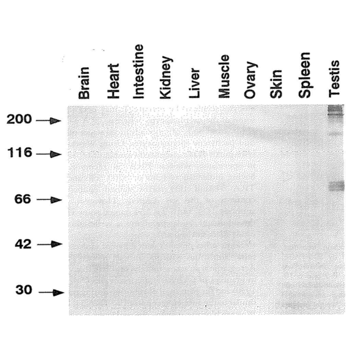
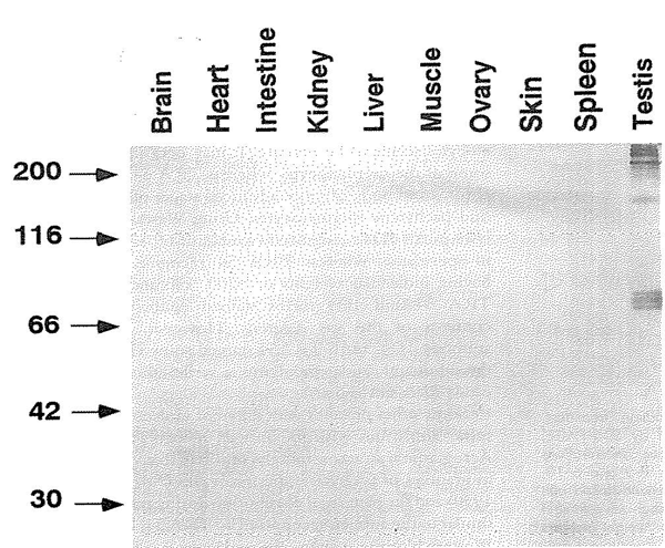
Many transcripts for proteins specifically expressed in haploid germ cells are synthesized in the spermatogenesis. A monoclonal antibody TRA54 recognizes specific organelles in germ cell cytoplasm from spermatocytes to spermatids (1, 2). Western blots using TRA54 revealed broad bands with 85 kD, 190 kD and >200 kDa in testis probably due to alternative splicing and/or post-translational modification (Fig. 1).
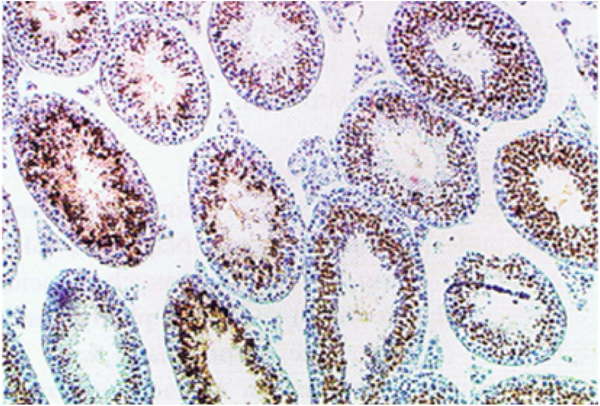
The molecular size diversity is due to the difference in glycosilation (Ref. 1). This antibody can be used for immunohistochemical staining of testicular tissues (1, 3). The hybridoma producing TRA54 was obtained from the immunaization of a rat with cell lysates of adult mouse testes by the group of Prof. Yoshitake Nishimune of Osaka University.
|
|
|
|
|
|
References (Antibody TRA54 has been used in the following publications.)
- Pereira LA et al “Characterization and expression of a stage specific antigen by monoclonal antibody TRA 54 in testicular germ cell” Int J Androl 21: 34-40 (1998) PMID: 9639150 WB, IHC
- Arrotéia KF et al “Identification and characterization of an antigen recognized by monoclonal antibody TRA 54 in mouse epididymis and vas deferens”J Androl 25: 914-921 (2004) PMID: 15477363 WB, IHC
- Ventelä S et al ”Intercellular organelle traffic through cytoplasmic bridges in early spermatids of the rat: mechanisms of haploid gene product sharing” Mol Biol Cell 14: 2768-2780 (2003) PMID: 12857863 IHC, Immno-Electron Microscopy
Aliases for SLA Gene
- Src Like Adaptor 2 3 5
- Src-Like-Adapter 2 3 4
- SLAP-1 2 3 4
- HSLAP 2 3 4
- SLAP 2 3 4
- Src-Like-Adapter Protein 1 3 4
- SLA1 2 3
- Src-Like-Adaptor 2
- SLAP1 4
- SLA 5
