


Anti-HB S Antibody Quantitative ELISA Kit (96assay)
Product#: FNK-BCL-AKS-01
$99,999,999.00
Availability:
Ships in 1-2 Weeks
Anti-HB S Antibody Quantitative ELISA Kit (96assay)
Cat. No.: FNK-BCL-AKS-01
Size: 1kit
Storage: 4℃
For inquiries about the price, please contact us at support@diagnocine,com
Introduction:
Three types of hepatitis B virus surface antigen (HBsAg), L-, M-, and S-proteins are known. The L-protein is composed of S, Pre-S2, and Pre-S1 domains, and the M-protein is composed of S, Pre-S2 domains, and the S-protein S domain alone. All three surface antigens have S antigen (S-protein) and detection of S antigen has been used for diagnosis of HBV infection because of its high content in serum of HBV-infected patient. The “HB-S Antigen Quantitative ELISA Kit, Rapid-II” can quantitatively detect the activity of S antigen.
The principle and outline of assay:
The key components of the kit are two types of anti-S antibody and the standard S antigen. So-called sandwich system is employed in this kit, where one type of antibody captures the antigen on the microplate surface, and the captured antigen is detected by another antibody labeled with HRP. Finally, the amount of HRP attached was determined using chromogenic substrate. There are two methods to detect S antigen using the kit, and the approximate times required, excluding times for sample preparation, plate washing, abs measurement etc, are as follows;
● 1-Step method: Total assay time = 90 min
①reaction with capture and detection IgGs(60) → ②chromogenic reaction(30)
● 2-Step method: Total assay time = 150 min
①reaction with capture IgG(60) → ②reaction with detection IgG(60) → ③chromogenic reaction(30)
[note] To get higher signal at low antigen concentration, increase reaction temperature to 37˚C. These procedures may cause high background and signal strength too high at higher STD concentration.
The definition of S antigen activity:
The strength of S antigen activities of L- and M-protein differs from that of S-protein presumably due to conformational difference. Therefore, in this kit S antigen activity was shown as unit system where 1 nUnit is defined as the activity expressed by 1 ng of the standard antigen that is provided in the kit. The one nUnit is roughly equal to 0.5 to 1.0 IU of WHO’s standard antigens.
Storage condition and stability:
All components can be stored at 4˚C. At the condition, all reagents are stable for at least 6 months from production date.
Kit composition:
The kit contains following materials. Please ensure that all materials are provided in the kit.
- Standard S Antigen (lyophilized form, recombinant antigen): 100 ng (1 tube)
- Buffer A, 20 x conc.: 20mL (1 bottle)
- Reaction buffer (3 x conc.): 20mL (1 bottle)
- Anti S Detection IgG (HRP-labeled): 12µL (1 tube)
- Chromogenic Reagent A: 6mL(1 bottle)
- Chromogenic Reagent B: 6mL (1 bottle)
- Stop Solution: 6.5 mL (1 bottle)
- Assay Plate For S Antigen: 1 plate (pre-coated with anti-S antigen antibody, strip type)
- User’s manual
Equipments required for the assay and Reagents not provided by the kit
- Microplate reader (equipped to measure absorbance at 450 nm)
- Micropipettes (for the handling of standard antigen and samples)
- Microtubes (for the handling of standard antigen and samples)
- Plate sealers (or plastic films)
- Multi (8-) channel pipette and its reservoir
- Distilled water or pure water
<Preparation before starting>
- Buffer A: Warm up Buffer A (20 x conc.) bottle to room temperature for complete dissolution (chilled concentrated buffer often contains depositions of salts), and dilute the content to 20-fold with distilled or pure water. This makes 400 mL of Buffer A (1 x conc.) ready to use. Storage of diluted Buffer A should be at 4˚C.
- Reaction Buffer: Warm up Reaction Buffer (3 x conc.) bottle to room temperature for complete dissolution (chilled concentrated buffer often contains depositions of salts), and dilute the content to 3-fold with distilled or pure water. This makes 60 mL of Reaction Buffer (1 x conc.). Storage of diluted reaction buffer should be at 4˚C.
- Standard antigen: Pour 1 mL of Buffer A into the standard antigen tube and invert it several times for complete dissolving. This makes 100nUnit/mL (100ng/mL) antigen solution.
1-Step Method
<①Preparation of standard solutions>
1. Prepare 7 new microtubes and print number from ① to ⑦. As indicated in the below table, transfer 900 μL of Reaction Buffer into No.1 microtube, add 100 μL of standard solution (100 nUnit/mL), and mix well. This makes 10 nUnit/mL of STD①. Likewise, prepare the series of standard solutions as indicated in below table.
2. To draw the standard curve, standard solution from STD① to STD⑦ are used.
3. Transfer 400 μL of Reaction buffer into a microtube as the blank.
| STD ID | Reaction buffer (or Buffer A)* |
Antigen solution for dilution | Final conc. | |||||||
| STD① | 900 | μL | + | 100 | nUnit/mL | 100 | μL | = | 10 | nUnit/mL |
| STD② | 500 | μL | + | 10 | nUnit/mL | 500 | μL | = | 5 | nUnit/mL |
| STD③ | 600 | μL | + | 5 | nUnit/mL | 400 | μL | = | 2 | nUnit/mL |
| STD④ | 500 | μL | + | 2 | nUnit/mL | 500 | μL | = | 1 | nUnit/mL |
| STD⑤ | 500 | μL | + | 1 | nUnit/mL | 500 | μL | = | 0.5 | nUnit/mL |
| STD⑥ | 800 | μL | + | 0.5 | nUnit/mL | 200 | μL | = | 0.1 | nUnit/mL |
| STD⑦ | 500 | μL | + | 0.1 | nUnit/mL | 500 | μL | = | 0.05 | nUnit/mL |
<②Preparation of sample solutions>
1. Dilute the sample solution by Reaction Buffer so that the expected activity is within the measurable range (0.05~10nUnit/mL). Sample should be diluted more than 10-fold by Reaction Buffer for accurate measurement.
2. When S antigen activity of a sample can not be estimated, please prepare multiple samples with different dilution factors.
3. Human serum sample should be diluted more than 100-fold by Reaction Buffer to prevent the effect of serum components.
<③One step reaction of antigen with capture IgG and detection IgG >
1. Take the assay plate out of the aluminum package.
2. In triplicate manner, add 100 μL of sample, standard and blank solutions to each well.
3. Dilute Anti S Detection IgG (HRP-labeled) to 500-fold with Reaction Buffer and transfer the solution to reservoir.
4. Add 50 μL of diluted Anti S Detection IgG into each well by using a 8-channel multi pipette.
5. Cover the plate by a plate sealer, and let it stay at room temperature (around 25°C) for 1 hr.
(go to “④Preparation of Chromogenic solution” for next step)
2-Step Method
<①Preparation of standard solutions>
Prepare standard solutions as indicated in “①Preparation of standard solutions“ for 1-Step Method. Use Buffer A for dilution instead of Reaction Buffer in 2-Step method.
<②Preparation of sample solutions>
Prepare sample solutions as indicated in “②Preparation of sample solutions“ for 1-Step Method. Use Buffer A for dilution instead of Reaction Buffer in 2-Step method.
<③ 2-Step reaction of antigen with capture IgG and detection IgG >
1. Take the assay plate out of the aluminum package.
2. In triplicate manner, add 100 μL of sample, standard solutions and blank to each well. Incubate at room temperature (around 25°C) for 1 hr.
3. Dilute Anti S Detection IgG (HRP-labeled) to 1000-fold with Reaction Buffer and transfer the solution to reservoir.
4. At the end of the reaction of sample with capture IgG, remove the reaction solution from each well by decantation followed by gentle tapping on the paper towel.
5. Wash each well with 300 μL/well of Buffer A three times and remove residual solution by gentle tapping on the paper towel.
6. Add 100μL of diluted Anti S Detection IgG (HRP-labeled) into each well by using 8-channel multi pipette, and incubate for 1 hr at room temperature (around 25°C).
<④Preparation of Chromogenic solution>
1. About 10 min before the end of reaction, prepare chromogenic reaction mixture as follows.
2. Transfer 6 mL of Substrate Solution A and 6 mL of Substrate Solution B into a reservoir for 8-channel pipette, and mix well.
[note] The prepared Chromogenic solution decreases the activity by oxidation within fairly short time. So please use the solution within 15 min after preparation.
<⑤Chromogenic reaction>
1. At the end of antibody reaction, remove the reaction solution from each well by decantation followed by gentle tapping on the paper towel.
2. Wash each well with 300 μL/well of Buffer A five times and remove residual solution by gentle tapping on the paper towel.
3. Add 100 μL of the Chromogenic solution to each well by using an 8-channel pipette, and incubate for 30 min at room temperature in a light shielded container.
4. Stop the reaction by adding 50 μL/well of Stop Solution.
[note] The blue color generated by Chromogenic reaction changes rather quickly. To obtain accurate determinations, it is important to measure the absorbance soon after the addition of stop solution.
<⑥Absorbance measurement and calculation>
1. Measure the absorbance at 450 nm of each well by using a plate reader.
2. Calculate the specific absorbance of each sample or standard by subtracting the blank value.
3. Calculate the best fit curve between the specific absorbance values and the corresponding standard concentrations using 4 parameters logistic model.
4. The concentration of samples can be calculated from the equation of the best fit curve. 5. The calculated concentrations which are out of the range (<0.05 or >10 nUnit/mL) are not reliable, and not used for determination.
[note] Clean up the bottom of the plate before absorbance measurement to avoid inaccurate measurement.
Example of standard curve
Below is an example of measurements of standard solutions and resulting standard curve.
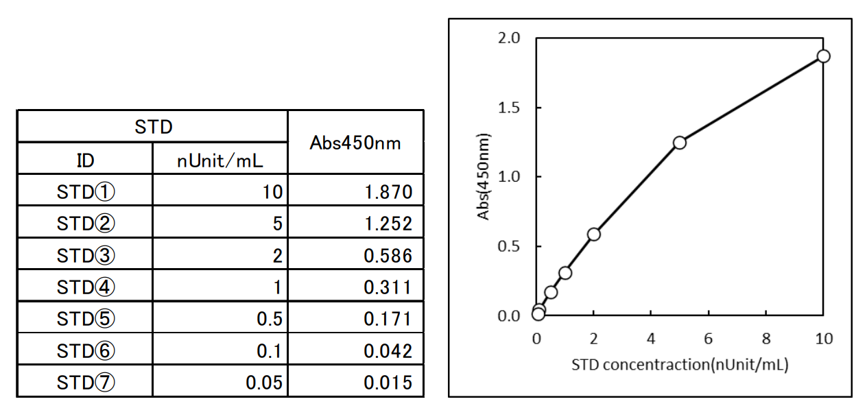
[note] The above data is an example obtained by 2-Step Method in the manufacturer's laboratory, and does not guarantee the performance of the kit. The results may differ due to the difference of the technique and other conditions.
Recently Viewed







Anti-MMAE, Mouse-Mono(AB12401)
$910.28
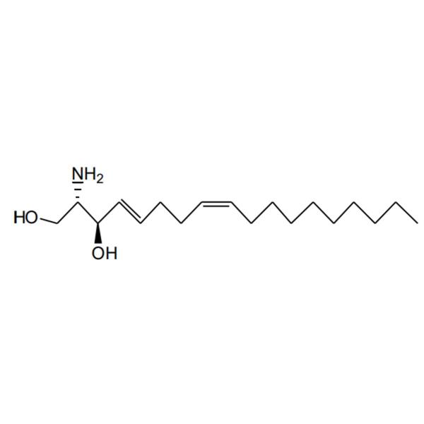
trans-4-cis-8-Sphingadienine
$404.58

MYBLOCK Mini Dry Bath
$66.71
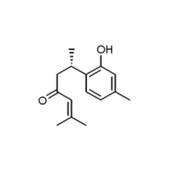
Turmeronol B
$606.86