Anti-CD40 antibody, mouse monoclonal (5C3), FITC conjugated
Anti-CD40 antibody, mouse monoclonal (5C3), FITC conjugated
General information
| Cat. No. | :FNK-72-032 |
| Size | :50 ug |
| Host Animal | :Mouse |
| Specificity | :Human |
| Isotype | :Mouse IgG1 k |
| Conjugate | :[FITC] / [IgG] = 5.5 |
| Label | :FITC |
| Clone | :5C3 |
| Immunogen | :Recombinant extracellular domain of CD40 |
| Application |
|
| Storage | :Shipped at 4℃ and stored at -20℃ or below |
| Shipping Condition | :Dry Ice |
| Form | :1.0 mg/ml in PBS, 50% glycerol, filter-sterilized |
| Data Link | :Swiss-Prot P25942 |
Description
CD40 is a 45-50-kDa glycoprotein belonging to the tumor necrosis factor (TNF) receptor superfamily. CD40 is specifically expressed on the surface of B cells and specialized antigen-presenting cells such as dendritic cells and macrophages. CD40 interacts with the CD40 ligand (CD154) which is found primarily on T cells, playing a role in both humoral and cell-mediated immune responses. Activation of CD40 on B cells by CD40 ligand causes B cell proliferation, differentiation, immunoglobulin isotype switching, germinal center formation, and stimulation of the humoral memory response.
This antibody reacts with a 45-48 kDa type I integral membrane glycoprotein present on peripheral blood and tonsillar B cells, but not expressed on terminally differentiated B cells.
The antibody against human CD40 was produced from a hybridoma (5C3) cultured in serum-free medium and purified under mild conditions by proprietary chromatography processes.
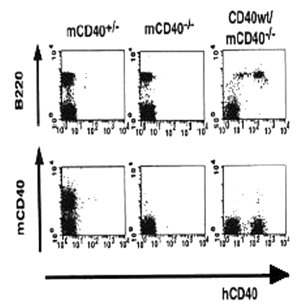 |
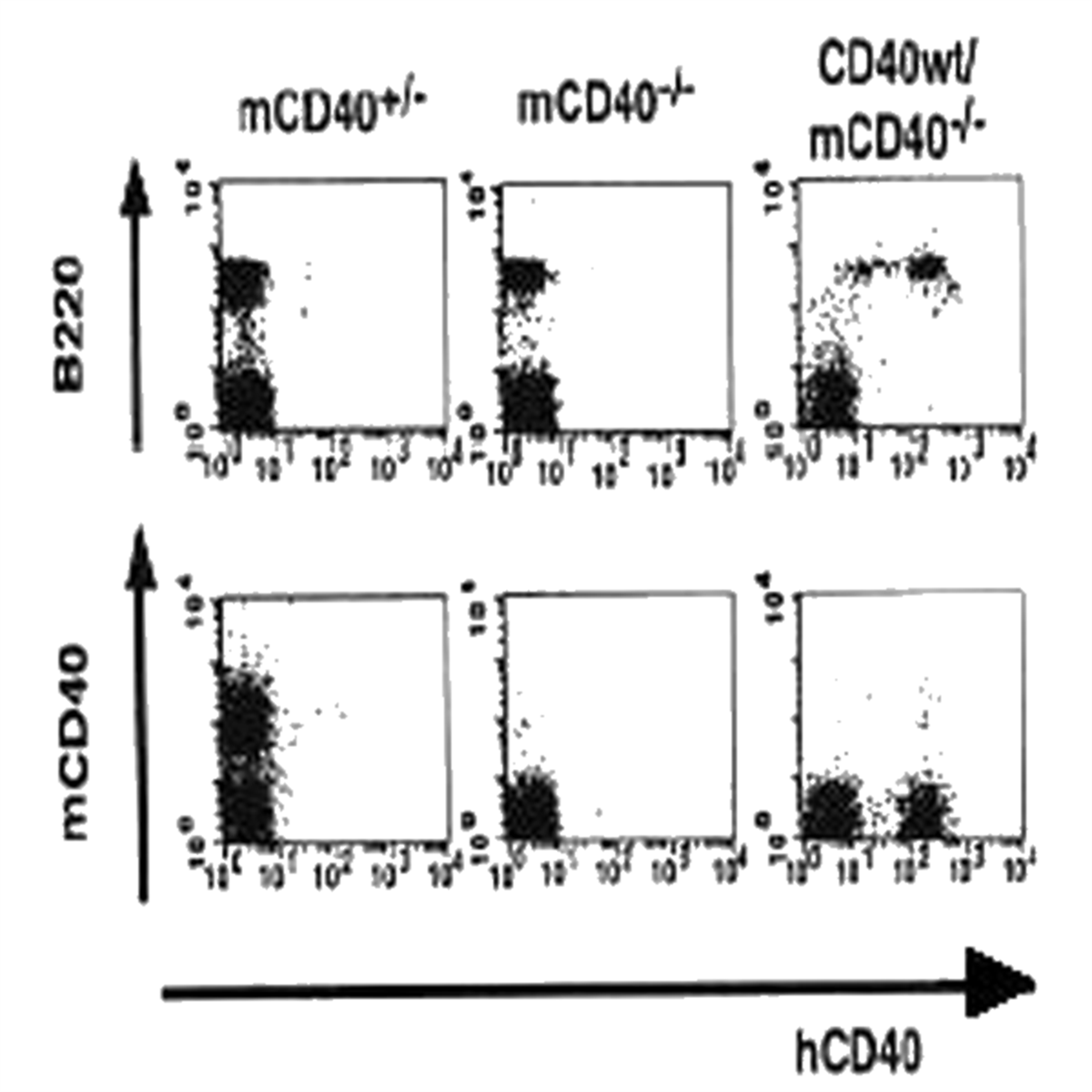
Fig.1 B cell-specific expression of h (human) CD40 in transgenic mice (ref.2). Splenocytes from m (mouse) CD40+/- , mCD40-/- and hCD40 wild type/mCD40-/- mice were stained with monoclonal antibodies against mCD40, B220 and hCD40 (5C3) and analyzed by flow cytometry. hCD40 molecules were expressed specifically on B220+ B cells |
Antigen Characterization
- Type: Primary
- Application: ELISA, Western Blot, Immunoprecipitation, Flow Cytometry, Immunohistochemistry
- Species: Human
- Recombinant: Yes
Human Diseases
- Hyper-IgM Immunodeficiency Syndrome Type 3 (HIGM3): A genetic disorder caused by defects in the CD40 gene, leading to an inability of B cells to undergo isotype switching and mount effective antibody responses.
- Autoimmune Diseases: Dysregulation of CD40 signaling can contribute to autoimmune conditions such as systemic lupus erythematosus (SLE) and rheumatoid arthritis (RA).
- Malignancies: Altered CD40 expression is implicated in various cancers, including B-cell lymphomas and solid tumors, affecting tumor immunity and progression.
Cellular Signaling Pathways
- CD40-CD40L Interaction: The binding of CD40 to its ligand (CD40L) on activated T cells is crucial for B cell activation, proliferation, and differentiation into memory B cells.
- NF-κB Pathway: CD40 signaling activates the NF-κB pathway, promoting survival and proliferation of B cells.
- MAPK Pathway: Activation of the MAPK pathway through CD40 engagement leads to the phosphorylation of transcription factors that enhance immune responses.
- PI3K/Akt Pathway: CD40 signaling can activate the PI3K/Akt pathway, which is involved in cell survival and growth.
Aliases for CD40 Gene
- CD40 Molecule 2 3 5
- Bp50 2 3 4
- Tumor Necrosis Factor Receptor Superfamily Member 5 3 4
- CD40 Molecule, TNF Receptor Superfamily Member 5 2 3
- CD40L Receptor 3 4
- TNFRSF5 3 4
- P50 2 3
- Tumor Necrosis Factor Receptor Superfamily, Member 5 2
- B Cell Surface Antigen CD40 3
- B-Cell Surface Antigen CD40 4
- B Cell-Associated Molecule 3
- CD40 Antigen 4
- CDW40 3
- CDw40 4
- CD40 5
References
- Inui S et al (1990) “Identification of the intracytoplasmic region essential for signal transduction through a B cell activation molecule, CD40.” Eur J Immunol 20: 1747-1753 PMID: 1698631
- Yasui T et al (2002) “Dissection of B cell differentiation during primary immune responses in mice with altered CD40 signals.” Int Immunol 14: 319-329 PMID: 11867568
- Ishida I et al (2003) “Involvement of CD100, a lymphocyte semaphoring, in the activation of the human immune system via CD72: implications for the regulation of immune and inflammatory responses.” Int Immunol. 15: 1027-1034 PMID: 12882840.
- Jodi L. Karnell a, et al. “Targeting the CD40-CD40L Pathway in Autoimmune Diseases: Humoral Immunity and Beyond.” Advanced Drug Delivery Reviews, Elsevier, 13 Dec. 2018, www.sciencedirect.com/science/article/pii/S0169409X18303089.
- Khalil, Magi, and Robert H Vonderheide. “Anti-CD40 Agonist Antibodies: Preclinical and Clinical Experience.” Update on Cancer Therapeutics, U.S. National Library of Medicine, 1 June 2007, www.ncbi.nlm.nih.gov/pmc/articles/PMC2706543/.
- Argiriadi, Maria A., et al. “CD40/Anti-CD40 Antibody Complexes Which Illustrate Agonist and Antagonist Structural Switches - BMC Molecular and Cell Biology.” BioMed Central, BioMed Central, 5 Aug. 2019, bmcmolcellbiol.biomedcentral.com/articles/10.1186/s12860-019-0213-4.
- Vonderheide, Robert H, and Martin J Glennie. “Agonistic CD40 Antibodies and Cancer Therapy.” Clinical Cancer Research?: An Official Journal of the American Association for Cancer Research, U.S. National Library of Medicine, 1 Mar. 2013, www.ncbi.nlm.nih.gov/pmc/articles/PMC3590838/.
- Xingx, et al. “CD40 Antibody (H-10): SCBT - Santa Cruz Biotechnology.” SCBT, 12 July 2018, www.scbt.com/p/cd40-antibody-h-10.