

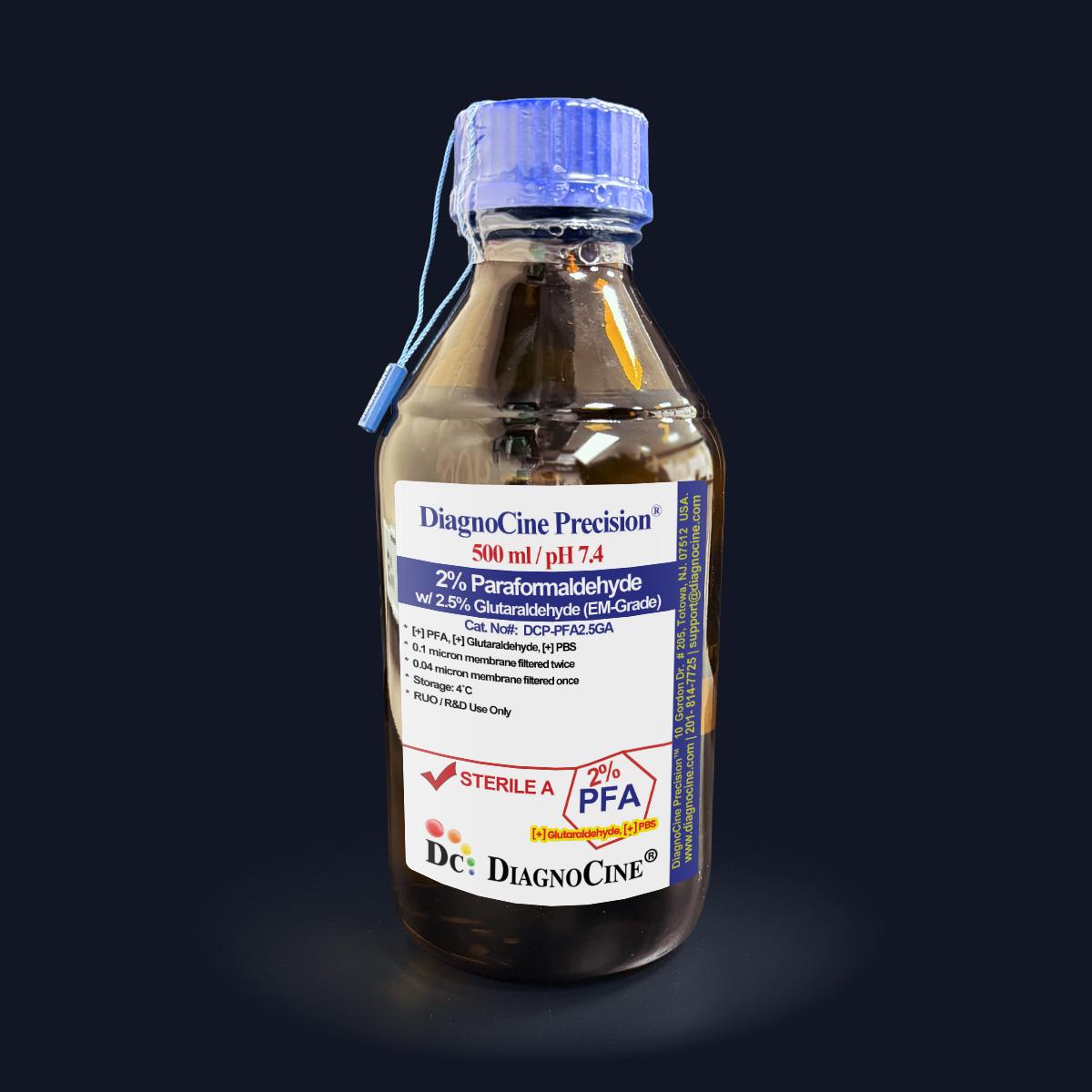
2% Paraformaldehyde with 2.5% Glutaraldehyde (EM-Grade)
FluxMPS™ 2% Paraformaldehyde with 2.5% Glutaraldehyde (EM-Grade)
A dual-aldehyde electron microscopy fixative combining 2% paraformaldehyde and 2.5% glutaraldehyde in a 0.1M phosphate buffer at pH 7.4. Formulated with EM-Grade PFA (≥ 99.9%) and EM-Grade Glutaraldehyde (Grade I), and processed through a quadruple-stage 0.1 µm and 0.04 µm membrane filtration system, this fixative is engineered to deliver rapid stabilization with thorough cross-linking for superior ultrastructural preservation.
- Dual-aldehyde formulation: 2% paraformaldehyde with 2.5% glutaraldehyde in 0.1M phosphate buffer
- Quadruple-stage filtration: 0.1 µm membrane filtered twice and 0.04 µm membrane filtered twice
- Physiological pH 7.4 buffering for optimal ultrastructural preservation
- Formulated with EM-Grade PFA (≥ 99.9%) and EM-Grade Glutaraldehyde (Grade I), plus EM-suitable salts (>98-100%)
- DNase and RNase activity tested — none detected after 18 hours at room temperature
- Manufactured under ISO 13485-certified, CE-approved facility conditions
- Supplied in amber, light-protected bottles — available in 500 mL and 1000 mL sizes
- Custom concentrations, pH, and additive formulations available on request — contact support@diagnocine.com
- pH
- 7.4
- Formulation
- 2% PFA / 2.5% Glutaraldehyde
- Buffer System
- 0.1M Phosphate Buffer
- Filtration
- 0.1 µm x2 + 0.04 µm x2
- DNase Activity
- None detected
- RNase Activity
- None detected
- Appearance
- Clear solution
- Storage
- 4°C, protected from light
- Shelf Life
- 12 months
- Shipping
- Typically within 48 hours
Engineered where standard fixatives fail
Conventional 0.22 µm-filtered fixative preparations can carry subvisible particulates, inconsistent pH, and undocumented raw-material grade — introducing artifacts into delicate ultrastructural preservation. FluxMPS™ buffers address each failure mode directly.
Microchannel-safe purity
Sequential 0.1 µm and 0.04 µm membrane filtration removes finer particulates than a single-pass 0.22 µm filter, supporting cleaner fixative preparations for electron microscopy imaging.
Precise, stable pH
Buffered at physiological pH 7.4 in 0.1M phosphate buffer for consistent, reproducible fixation chemistry across specimens.
Ultrapure-grade water
Manufactured using Ultrapure Type 1 water (18.2 MΩ·cm), consistent with USP <85> production standards.
Low-artifact ultrastructural preservation
The dual-aldehyde mechanism preserves antigenic sites for immunohistochemistry while delivering excellent ultrastructural detail for electron microscopy.
Defined, traceable composition
Formulated with EM-Grade PFA (≥ 99.9%) and EM-Grade Glutaraldehyde (Grade I), with DNase and RNase activity confirmed absent on every lot.
Customization on demand
Alternate concentrations, pH, and additions of chemicals, compounds, proteins, or supplements are available — contact support@diagnocine.com.
Quadruple-stage filtration system
This fixative is processed through a four-stage membrane filtration sequence — 0.1 µm membrane filtration twice and 0.04 µm membrane filtration twice — supporting a cleaner preparation for demanding electron microscopy and immunohistochemistry workflows.
-
1
0.1 µm Pre-filtration I
Removes large particulates and aggregates, extending the service life of downstream filters.
-
2
0.04 µm Pre-filtration II
Retains fine particulates and bioburden ahead of terminal filtration.
-
3
0.1 µm Sterile-filtration I
A second 0.1 µm pass provides redundant particulate control.
-
4
0.04 µm Sterile-filtration II — Final Polish
A final 0.04 µm polish helps reduce mycoplasma-scale contaminants, since the smallest mycoplasma species measure about 0.2 microns in diameter.
Performance vs. conventional buffer
Sequential 0.1 µm and 0.04 µm filtration removes finer particulates than a single 0.22 µm pass typically used in conventional fixative preparations.

© Diagnocine® — DCP-PFA2.5GA
Where dual-aldehyde fixation delivers
This fixative rapidly stabilizes cellular ultrastructure through paraformaldehyde penetration, followed by durable glutaraldehyde cross-linking — supporting a broad range of specimen types and imaging modalities.
Automated Bioreactors & Robotics
For automated fixation and liquid-handling platforms, an optional 0.01 µm (10 nm) ultra-filtered variant of this fixative can be requested to further protect fine-tolerance fluidic components.
- Total Particulate Exclusion for automated dispensing lines
- Valve & Sensor Protection in robotic liquid handlers
- Extended Perfusion Stability during automated fixation cycles
Inquiry Required: the 0.01 µm ultra-filtered grade is available on request — contact support@diagnocine.com.
Micro Physiological System (MPS) & Chip Fixation
Fixation of on-chip tissue and organ models for downstream structural and ultrastructural analysis.
Specimen Fixation & Wash Processing
Immersion or perfusion fixation followed by the recommended 3 × 5 minute wash in fixation buffer prior to further processing.
iPSC-Derived Model Handling
Structural preservation of iPSC-derived cell models while maintaining antigenic sites for downstream immunolabeling.
Endothelial & Primary Cell Perfusion
Perfusion fixation of vascular and primary cell preparations for ultrastructural and morphological assessment.
Immunohistochemistry Studies
Preserves antigenic sites for immunohistochemical detection while stabilizing overall tissue architecture.
Electron & Optical Microscopy
Delivers excellent ultrastructural preservation for electron microscopy specimen preparation and related optical imaging workflows.
Full product specifications
All parameters below reflect the measured and stated values for DCP-PFA2.5GA as provided by Diagnocine quality control.
| Parameter | Specification |
|---|---|
| Formulation / Composition | 2% Paraformaldehyde, 2.5% Glutaraldehyde, 0.1M Phosphate Buffer |
| Appearance | Clear solution |
| pH USP <791> | 7.4 |
| Buffer System | 0.1M Phosphate Buffer |
| Parameter | Specification |
|---|---|
| Terminal Filtration | 0.1 µm membrane twice and 0.04 µm membrane twice |
| Raw Fixative Pre-filtration (Non-Sterile stage) | 0.02 µm membrane once and 0.01 µm membrane once |
| DNase Activity | None detected (18 hours, room temperature, plasmid DNA) |
| RNase Activity | None detected (18 hours, room temperature, ribosomal RNA) |
| Manufacturing Standard ISO 13485 | ISO 13485-controlled conditions |
| Parameter | Specification |
|---|---|
| Storage Temperature | 4°C, away from bright light (amber bottle) |
| Aliquot Storage (-20°C, amber, near-zero air space) | 1 year |
| Unopened Storage (4°C) | 4 months |
| Open / Routine Use Storage (4°C) | 2 weeks |
| Shelf Life | 12 months |
| Shipping Condition | Ships typically within 48 hours; may take up to 1 week during unforeseen events |
| Post-fixation Specimen Storage | 4°C for up to one week before further processing |
| Parameter | Specification |
|---|---|
| Raw Material Grade | EM-Grade PFA (≥ 99.9%), EM-Grade Glutaraldehyde (Grade I), EM-suitable salts (>98-100%) |
| Final Packaging Notice | Amber bottle; not vacuum-sealed or nitrogen-purged; not certified as an EM-grade preparation; EM-grade certificate will not be issued |
| Manufacturing QMS ISO 13485 | ISO 13485-certified and CE-approved facilities (DiagnoCine Precision); final packaging, QA, and testing at the DiagnoCine R&D and Quality Testing Center |
| Assembly Location | Customization requests and assembly completed at DiagnoCine Precision, Totowa, New Jersey, USA |
| Intended Use | Fixative for electron microscopy (EM) / suitable for EM use; For Research Use Only |
Full composition
This dual-aldehyde fixative is formulated in 0.1M phosphate buffer at physiological pH for optimal specimen preservation.
| Component | CAS Number | Concentration |
|---|---|---|
| Paraformaldehyde (PFA) | 30525-89-4 | 2% |
| Glutaraldehyde | 111-30-8 | 2.5% |
| Phosphate Buffer | 0.1M |
Manufacturing & compliance
DCP-PFA2.5GA is manufactured and packaged under controlled, documented conditions to support consistent, reproducible fixation performance.
ISO 13485:2016 QMS
Manufactured under ISO 13485-certified and CE-approved facility conditions (DiagnoCine Precision).
Ultrapure Type 1 Water
Formulated using Ultrapure Type 1 water (18.2 MΩ·cm), aligned with USP <85> production practice.
Fill & Packaging Notice
Supplied in an amber bottle; the final solution is not vacuum-sealed or nitrogen-purged and is not certified as an EM-grade preparation. An EM-grade certificate will not be issued.
Micro-Batch Precision
Final packaging, quality assurance, and testing completed at the DiagnoCine R&D and Quality Testing Center; customization and assembly performed at DiagnoCine Precision, Totowa, New Jersey, USA.
DNase Activity
None detected after incubation of plasmid DNA with this product for 18 hours at room temperature.
RNase Activity
No RNase activity detected after incubation of ribosomal RNA with this product for 18 hours at room temperature.
pH Verification
Verified at pH 7.4 as part of release quality control testing.
Documentation / CoA
Contact support@diagnocine.com for available lot documentation.
How DCP-PFA2.5GA compares
A side-by-side comparison against conventional EM fixative preparations.
| Parameter | DCP-PFA2.5GA (FluxMPS™) | Conventional EM Fixative (0.22 µm filtered) | Standard Alternative (0.22 µm filtered) |
|---|---|---|---|
| Formulation type | Dual-aldehyde (2% PFA + 2.5% Glutaraldehyde) | Single-aldehyde or undefined ratio | Single-aldehyde or undefined ratio |
| Final filtration pore size | 0.04 µm | 0.22 µm | 0.22 µm |
| Number of filtration stages | 4 | 1 | 1 |
| DNase / RNase tested | check_circle | cancel | cancel |
| EM-grade raw components | check_circle | cancel | cancel |
| ISO 13485-certified manufacturing | check_circle | cancel | cancel |
| Amber, light-protected packaging | check_circle | cancel | cancel |
| Physiological pH buffering (0.1M phosphate, pH 7.4) | check_circle | cancel | cancel |
| Custom concentration / pH formulation available | check_circle | cancel | cancel |
| EM-grade certification issued | cancel | cancel | cancel |
Frequently asked questions
Common questions about DCP-PFA2.5GA formulation, filtration, and handling.
Supporting literature
Curated literature relevant to dual-aldehyde fixation chemistry and electron microscopy specimen preparation.
- Karnovsky, M.J. A formaldehyde-glutaraldehyde fixative of high osmolality for use in electron microscopy. J Cell Biol. doi:10.1083/jcb.27.2.137a
- Hopwood, D. Fixation and fixatives: a review. Histochem J. doi:10.1007/BF01004606
- Hayat, M.A. Principles and techniques of electron microscopy: biological applications. Cambridge University Press. doi:10.1017/CBO9780511812255
- Fox, C.H. et al. Formaldehyde fixation. J Histochem Cytochem. doi:10.1177/33.8.3894502
- Kiernan, J.A. Formaldehyde, formalin, paraformaldehyde and glutaraldehyde: what they are and what they do. Microsc Today. doi:10.1017/S1551929500057060
- Bozzola, J.J., Russell, L.D. Electron microscopy: principles and techniques for biologists. Jones and Bartlett. doi:10.1002/jemt.1060030110
- Eltoum, I. et al. Introduction to the theory and practice of fixation of tissues. J Histotechnol. doi:10.1179/his.2001.24.3.173
- Ushiki, T. Collagen fibers, reticular fibers and elastic fibers: a comprehensive understanding from a morphological viewpoint. Arch Histol Cytol. doi:10.1679/aohc.65.109