

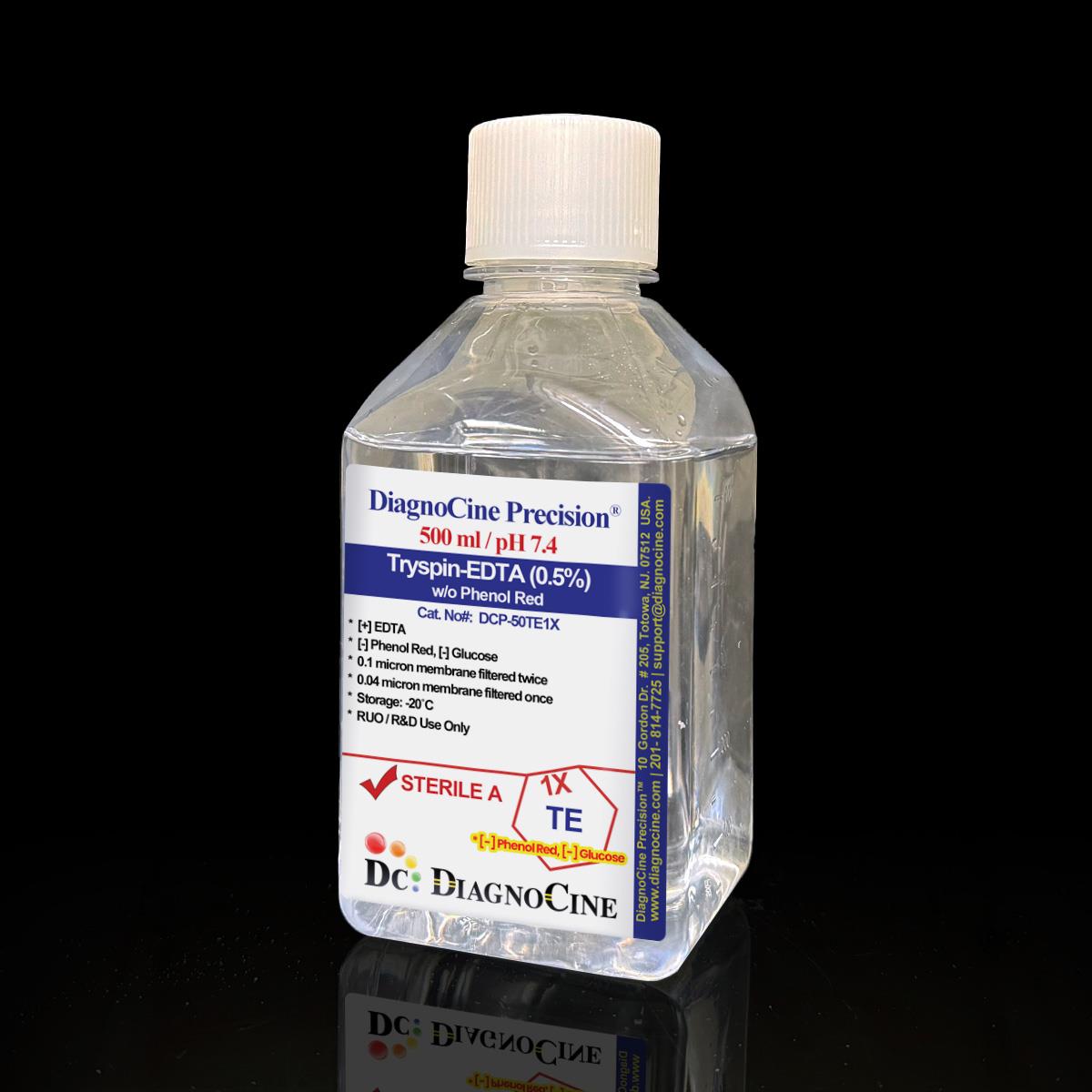
Trypsin (0.5%) with EDTA without Phenol Red (1X)
FluxMPS™ Trypsin (0.5%) with EDTA without Phenol Red (1X)
Microfluidics Suitable, quadruple-stage ultra-filtered (0.1 µm ×2 + 0.04 µm ×2) 1X Trypsin (0.5%)/EDTA solution engineered for rapid, reliable dissociation of strongly adherent cells, transformed fibroblasts, multilayer cultures and select tumor lines. Formulated without phenol red or glucose for a clean, customizable dissociation step upstream of sensitive imaging, metabolic and microfluidic / organ-on-a-chip (OoC) workflows.
- Ultra-filtered 0.5% Trypsin / 1.1 mM EDTA dissociation reagent, quadruple-stage filtered to a 0.04 µm final cut-off (0.1 µm ×2 + 0.04 µm ×2)
- Phenol red-free and glucose-free formulation for low-background microscopy and downstream assays sensitive to residual carbohydrate or dye carryover
- Formulated in a balanced PBS salt solution; EDTA chelates calcium to weaken cell-matrix adhesion and enhance trypsin's proteolytic detachment of strongly adherent cells
- Rapid detachment typically within 5 minutes at 37°C — markedly faster than collagenase-based dissociation, which can require up to 60 minutes
- Tested and confirmed free of detectable DNase and RNase activity after 18-hour incubation at room temperature
- Manufactured under an ISO 13485:2016 quality management system; final packaging, QC and customization completed at Diagnocine, Totowa, NJ
- Custom formulations available on request, including addition of D-glucose (dextrose) for applications requiring a carbon source during dissociation
- Trypsin0.5%
- EDTA1.1 mM
- Phenol RedNot added
- GlucoseNot added
- pH (USP <791>)7.4
- AppearanceClear liquid
- Filtration0.1 µm ×2 + 0.04 µm ×2
- Storage-20°C, long-term
- Shelf Life24 months from manufacture, unopened
- ShippingDry ice
Engineered where standard dissociation reagents fall short
Conventional 0.22 µm-filtered trypsin/EDTA solutions can carry sub-micron particulates and mycoplasma-scale contaminants into sensitive microfluidic channels, imaging systems and downstream chip cultures. FluxMPS™ is built to a tighter standard from the first filtration pass.
Microchannel-safe purity
Quadruple-stage filtration down to a 0.04 µm final pore size reduces sub-micron particulates that can accumulate in narrow microfluidic channels and valves during cell harvest and reseeding.
Defined, customizable formulation
Supplied without phenol red or glucose so the dissociation step does not introduce dye carryover or an undefined carbon source into downstream assays. D-glucose can be added back on request.
Ultrapure processing water
Formulated with ultrapure, low-bioburden water as part of the multi-stage filtration process, reducing trace contaminants that can interfere with sensitive cell-based assays.
Low background for imaging
Phenol red-free formulation minimizes optical interference during confocal microscopy and fluorescence-based assays performed shortly after cell detachment.
Fast, consistent detachment
0.5% trypsin combined with 1.1 mM EDTA in PBS is formulated for rapid dissociation of strongly adherent cells, transformed fibroblasts and multilayer cultures, typically within 5 minutes at 37°C.
Customization on demand
Alternate trypsin concentrations, added D-glucose/dextrose, alternate pH, and other chemical/compound additions are available — contact support@diagnocine.com.
Quadruple-stage filtration system
This Trypsin (0.5%)/EDTA reagent is processed through a validated four-pass filtration train reaching a 0.04 µm final pore size — two dedicated prefilter-plus-final-filter pairs run in series for full redundancy.
- 1
0.1 µm Prefiltration I
Removes large particulate, cell debris and protein aggregates, protecting the first 0.04 µm final filter cartridge.
- 2
0.04 µm Final filtration I
First 0.04 µm pass; retains sub-micron particulates and microaggregates that a standard 0.22 µm filter would pass.
- 3
0.1 µm Prefiltration II
Second dedicated prefilter, protecting the second 0.04 µm cartridge downstream.
- 4
0.04 µm Final filtration II — Polish
Ultimate polishing filter prior to aseptic fill & finish.
Performance vs. conventional media
Each 0.04 µm final filter is protected by its own dedicated 0.1 µm prefilter, giving two complete prefilter-plus-final-filter passes rather than a single descending cascade.

© Diagnocine® — DCP-50TE1X
Where fast, defined dissociation matters
Designed for strongly adherent cells, transformed fibroblasts, multilayer cultures and select tumor cell lines that resist gentle dissociation methods, across conventional and microfluidic culture formats.
Automated Bioreactors & Robotics
For automated perfusion bioreactors and liquid-handling robotics where residual particulate can foul valves and sensors, an optional 0.01 µm (10 nm) ultra nano-filtered MPS Grade variant of this reagent is available on inquiry.
- Total Particulate Exclusion — six-stage cascade down to 0.01 µm for automated systems
- Valve & Sensor Protection — reduces fouling risk in closed-loop perfusion hardware
- Extended Perfusion Stability — supports longer unattended run times between service intervals
Inquiry Required: The 0.01 µm (MPS Grade) variant is manufactured to order — contact support@diagnocine.com to discuss requirements.
Micro Physiological System (MPS) & Chip Harvest
Used to detach cells from microfluidic channels and chip substrates prior to reseeding or downstream analysis.
Passaging Strongly Adherent Tumor Lines
Higher trypsin concentration aids detachment of dense, matrix-rich tumor cell cultures during routine passaging.
iPSC-Derived Model Handling
May be used judiciously for dissociation steps where rapid detachment is prioritized; evaluate cell-surface protein sensitivity per cell type.
Endothelial & Primary Cell Passaging
Formulated to balance rapid detachment with viability for routine passaging of strongly adherent primary and endothelial cultures.
Pre-Assay Cell Harvest
Glucose-free formulation avoids introducing an undefined carbon source immediately before metabolic flux sampling or tracer studies.
Microscopy & Optical Sensing Workflows
Phenol red-free formulation reduces optical background during confocal imaging and biosensor readouts performed shortly after harvest.
Full specification sheet
Key physical, chemical, sterility and logistics parameters for DCP-50TE1X.
| Parameter | Specification |
|---|---|
| Formulation | EDTA present (chelating agent); phenol red not added; glucose not added |
| Appearance | Clear liquid |
| pH USP <791> | 7.4 |
| Trypsin concentration | 0.5% |
| EDTA concentration | 1.1 mM |
| Sodium Chloride | 137 mM |
| Potassium Chloride | 2.7 mM |
| Phosphate buffer (Na2HPO4 / KH2PO4) | 8.06 mM / 1.47 mM |
| Parameter | Specification |
|---|---|
| Sterility | Achieved via quadruple-stage 0.1 µm / 0.04 µm filtration (0.1 µm ×2 + 0.04 µm ×2) |
| Mycoplasma | 0.1 µm mycoplasma-retentive filtration (not tested per lot) |
| DNase activity | None detected (18 hr, room temperature, plasmid DNA) |
| RNase activity | None detected (18 hr, room temperature, ribosomal RNA) |
| Parameter | Specification |
|---|---|
| Storage | -20°C, long-term |
| Shelf life | 24 months from date of manufacture, unopened |
| Shipping condition | Dry ice |
| Parameter | Specification |
|---|---|
| Raw material grade | Ultrapure reagent-grade components |
| Traceability | Lot-based; final QC at Diagnocine R&D and Quality Testing Center, Totowa, NJ |
| Manufacturing QMS | ISO 13485:2016 |
| UNSPSC | 41116155 · Molecular biology and cell culture growth media · UNv260801 |
| Regulatory alignment | ISO 13485:2016 |
| Intended use | For Research Use Only (RUO) |
Full composition (mg/L equivalent / mM)
Total: 6 components, released per manufacturing lot. Custom concentrations, pH and additions (including D-glucose) available on request.
| Component | CAS Number | Concentration |
|---|---|---|
| Sodium Chloride (NaCl) | 7647-14-5 | 137 mM |
| Potassium Chloride (KCl) | 7447-40-7 | 2.7 mM |
| Sodium Phosphate Dibasic (Na2HPO4) | 7558-79-4 | 8.06 mM |
| Potassium Phosphate Monobasic (KH2PO4) | 7778-77-0 | 1.47 mM |
| Component | CAS Number | Concentration |
|---|---|---|
| No amino acid components in this formulation. | ||
| Component | CAS Number | Concentration |
|---|---|---|
| Trypsin | 9002-07-7 | 0.5% |
| EDTA | 60-00-4 | 1.1 mM |
Manufacturing & compliance
Manufactured under an ISO 13485-certified quality management system, with final packaging, QC and customization completed at Diagnocine.
DNase / RNase Activity
None detected after 18-hour incubation with plasmid DNA / ribosomal RNA at room temperature.
Sterility
Achieved via quadruple-stage 0.1 µm / 0.04 µm filtration performed in a sterile environment.
pH USP <791>
Released at pH 7.4.
Documentation / CoA
Certificate of Analysis available on request, documenting lot-specific QC results.
- Appearance, pH, and clarity
- Sterility (quadruple-stage filtration)
- DNase and RNase activity
How DCP-50TE1X compares
FluxMPS™ DCP-50TE1X against conventional 0.22 µm-filtered trypsin/EDTA dissociation reagents.
| Parameter | DCP-50TE1X (FluxMPS™) | Conventional trypsin-EDTA (0.22 µm filtered) | Standard alternative (0.22 µm filtered) |
|---|---|---|---|
| Grade | Microfluidics Suitable | Not specified | Not specified |
| Formulation | Phenol red-free, glucose-free | Often contains phenol red | Often contains phenol red |
| Final filtration pore size | 0.04 µm | 0.22 µm | 0.22 µm |
| Number of filtration stages | 4 | 1 | 1 |
| Mycoplasma barrier filtration | check_circle | cancel | cancel |
| Endotoxin (release specification) | FluxMPS™ — Not specified | Corning classical liquid media — < 0.25 EU/mL Sigma-Aldrich DMEM complete medium — ≤ 2 EU/mL Gibco classical DMEM — Not specified (recorded per lot) |
|
| Manufacturing QMS | ISO 13485:2016 | Varies by supplier | Varies by supplier |
| Microfluidic / OoC harvest compatibility | check_circle | Varies | Varies |
| Custom formulation | check_circle | cancel | cancel |
Comparison figures from published supplier specifications, accessed 2026-09-02. Suppliers that publish no numeric endotoxin specification are shown as "Not specified".
Frequently asked questions
Common questions about DCP-50TE1X Trypsin (0.5%) with EDTA without Phenol Red.
Supporting literature
Curated references on trypsin-EDTA cell dissociation, mycoplasma control by filtration, and microfluidic / organ-on-a-chip cell handling.
- Huang, H.-L. et al. Trypsin-induced proteome alteration during cell subculture in mammalian cells. J. Biomed. Sci. 17, 36 (2010). doi:10.1186/1423-0127-17-36
- Tsuji, K. et al. Effects of trypsin-EDTA treatment on cell surface proteins and adhesion molecule expression. Cytotechnology 68, 1701–1710 (2016). doi:10.1007/s10616-015-9927-0
- Bahmad, H.F. et al. Modeling adherent cell culture dissociation for downstream single-cell applications. Front. Oncol. 8, 347 (2018). doi:10.3389/fonc.2018.00347
- Bhatia, S.N. & Ingber, D.E. Microfluidic organs-on-chips. Nat. Biotechnol. 32, 760–772 (2014). doi:10.1038/nbt.2989
- Zhang, B. & Radisic, M. Organ-on-a-chip devices advance to market. Lab Chip 17, 2395–2420 (2017). doi:10.1039/C6LC01554A
- Uphoff, C.C. & Drexler, H.G. Detecting mycoplasma contamination in cell cultures by polymerase chain reaction. Methods Mol. Biol. 731, 93–103 (2011). doi:10.1007/978-1-61779-080-5_8
- Nikfarjam, L. & Farzaneh, P. Trypsinization and its effects on cell surface receptors. Cell J. 14, 155–158 (2012).
- Ivanovska, J. et al. Biocompatibility of EDTA-based chelation for cell dissociation in tissue engineering applications. Materials 13, 5299 (2020). doi:10.3390/ma13235299
- Sonnaert, M. et al. Quantitative validation of the presto blue metabolic assay for tissue-engineered constructs. Tissue Eng. Part C 21, 519–529 (2015). doi:10.1089/ten.tec.2014.0464
It works very well.?
Csanad *****
I like the product, I use it in flow cytometry; I just add a buffer after trypsinization, and run the cells without centrifuging. It works very well.
I also appreciate the fast shipping.