


Streptolysin O
Streptolysin O
Cat.No: FNK-01-531Size: 20 µg
Storage: -20℃, -80℃ (for longer storage) Aviod freeze-thaw cycles
Concentration: 1.0 mg/ml
Buffer: PBS (-), 1 mM DTT, 50% glycerol, sterilized by filtration. No additive nor carrier protein.
Purity: Over 98% by SDS-PAGE (see Fig.1)
Application
1. Functional studies2. Reagent for membrane pore formation to introduce small-to-macromolecules into living cells (Ref.1)
3. Antigen for the measurement of anti-streptolysin O antibody (ASO) (diagnostic reagent), ELISA
4. Western blotting, Dot blotting, SDS-PAGE
5. Immuno-chromatography
Background
Streptolysin O (SLO) is a membrane-damaging extracellular toxin produced by hemolytic streptococci. The membrane-damaging activity is measured by hemolysis of red-blood cells. SLO is easily inactivated in the presence of oxygen but can be reactivated by thiol compounds, so it is also called thiol-activated cytolysin. SLO is produced not only by Group A hemolytic streptococci but also by Group C and Group G strains. The amino acid sequences are highly conserved among them and their homology is over 98%.Health Hazard Data
LD50 - Lethal dose (50 percent kill) intravenous,
Rabbit: 1500 ng/kg (Ref:PHTHDT Pharmacology and Therapeutics. (Pergamon Press Ltd.,Headington Hill Hall, Oxford OX3 0BW, UK) Vol.(Issue) 11, Page 661,1981 )
Guinea pig: 12 µg/kg (Ref: BICMBE Biochimie.(SPPIF, B.P.22, F-41353 Vineuil, France, Vol.(Issue)55,Page 1187, 1973)
Toxicity is much less when introduced via other routes of entry like Interdermal injection.
Data Link
UniProtKB Q5PY51 (TACY_STREQ)
Please Note: All products are FOR RESEARCH USE ONLY. NOT FOR USE IN DIAGNOSTIC PROCEDURES. NOT FOR MILITRAY USE.
Data Images
01-531 Streptolysin O (Hemolytic streptococcus)
Fig.1 Purified SLO analysed by SDS-Page
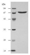 |
The SLO has molecular mass of 60.4 kDa. This product has melcular mass of 64.5 kDa. |
Fig.2 Introduction of fluorescein dextran of different molecular weights into resealed cells
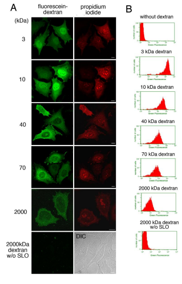 |
A. HeLa cells were incubated with or without (2000 kDa dextran w/o SLO) 0.13 µg/ml SLO on ice for 5 min. After wash with PBS three times, the cells were further with transport buffer containing propidium iodide at 32°C for 5 min. Semi-intact HeLa cells were incubated with 1.5 mg/ml L5178Y cytosol, an ATP regenerating system, GTP, glucose, and 100 µg/ml fluoresceindextran of 3, 10, 40, 70, or 2000 kDa at 32°C for 15 min, and then were resealed by treatment with 1 mM CaCl2 at 32°C for 5 min. After incubation with DMEM supplemented with FCS for 30 min, the cells were observed by confocal microscopy. Since the cells without SLO treatment did not contain the fluorescence of propidium iodide, differential interference contrast (DIC) image was shown. Bar = 10 µm. B. HeLa cells were treated as described in A, were trypsinized, and were subjected to flowcytometry. The histograms of fluorescein fluorescence of dextran with different molecular weight in PI-positive cells were shown. Data from Kano F. et al, PLoS One. 2012;7(8):e44127. |
References
1. Walev, I. et al. “Delivery of proteins into living cells by reversible membrane permeabilization with streptolysinO.” PNAS 98: 3185-3190 (2001) PMID: 11248053 Reagent for membrane pore formation to introduce small-tomacromolecules into living cells
2. Palmer, M. “The family of thiol-activated, cholesterol-binding cytolysins.” Toxicon 39: 1681-1689 (2001) PMID:11595631
3. Maeda, Y. et al. GPHR is a novel anion channel critical for acidification and functions of the Golgi apparatus. Nat. Cell Biol. 10: 1135-45 (2008) PMID: 18794847 Permeabilization of cells.
4. Furukawa K. et al. Reduction-triggered fluorescent amplification probe for the detection ofendogenous RNAs in living human cells. Bioconjug Chem. 2009 May 20;20(5):1026-36. PMID: 19374406 Introduction of probes for RNA into permeabilized human HL cells.
1. Thiery J. et al. Perforin activates clathrin- and dynamin-dependent endocytosis, which is required for plasma membrane repair and delivery of granzyme B for granzyme-mediated apoptosis. Blood 2010 115:1582-1593. PMID: 20038786 Promotion of endocytosis.
2. Kano F. et al. Hydrogen peroxide depletes phosphatidylinositol-3-phosphate from endosomes in a p38 MAPKdependent manner and perturbs endocytosis. Biochim Biophys Acta. 2011 May;1813(5):784-801. PMID: 21277337. Permeabilization of HeLa cells.
3. Potez S. et al. Tailored protection against plasmalemmal injury by annexins with different Ca2+ sensitivities. J Biol Chem. 2011 May 20;286(20):17982-91. PMID:21454475 Permeabilization of HEK cells.
4. Kano F. et al. A resealed-cell system for analyzing pathogenic intracellular events: perturbation of endocytic pathways under diabetic conditions. PLoS One. 2012;7(8):e44127. PMID: 22952896 Introduction of molecules into HeLa cells.
5. Imai A. et al. MADD/DENN/Rab3GEP functions as a guanine nucleotide exchange factor for Rab27 during granule exocytosis of rat parotid acinar cells. Arch Biochem Biophys. 2013 Aug 1;536(1):31-7. PMID: 23702376. Introduction of antibody into cell.
6. Gao N and Lehrman MA. Mannose-6-Phosphate: A Regulator of LLO Destruction. : Inka Brockhausen (ed.), Glycosyltransferases: Methods and Protocols, 2013, Springer, Methods in Molecular Biology, vol. 1022, DOI 10.1007/978- Inka Brockhausen (ed.), Glycosyltransferases: 1-62703-465-4_20. Link:.springer.com/protocol/10.1007 Introduction of mannose-6-phosphate into living cells. (The authors specifically recommend BioAcdemia streptolysin O for cell permeabilization)
7. Matsuto M et al. Reconstitution of the targeting of Rab6A to the Golgi apparatus in semi-intact HeLa cells: A role of BICD2 in stabilizing Rab6A on Golgi membranes and a concerted role of Rab6A/BICD2 interactions in Golgito-ER retrograde transport. Biochim Biophys Acta. 2015 Oct;1853(10 Pt A):2592-609. PMID: 25962623 Introduction of protein (Rab6A) into permeabilized Hela cells.
8. Yasuga H. et al. Logic gate operation by DNA translocation through biological nanopores. PLoS One. 2016 Feb 18;11(2):e0149667. PMID: 26890568 Nanopore formation in bilayer lipid menmbrae.
9. Ojima K. et al. Myosin substitution rate is affected by the amount of cytosolic myosin in cultured muscle cells. Anim Sci J. 2017 Nov;88(11):1788-1793. PMID:28631391 Permeabilization of cells
10. Kano F. et al. Establishment and phenotyping of disease model cells created by cell-resealing technique. Sci Rep. 2017 Nov 9;7(1):15167. PMID:2912317011. Reversible permeabilization of plasma membrane
11. Watanabe H. et al. Analysis of Pore Formation and Protein Translocation Using Large Biological Nanoporores. Anal Chem. 2017 Nov 7;89(21):11269-11277. PMID:28980803. Permeabilization for protein translocation.
Related Products
| SKU# | Description | Link |
|---|---|---|
| FNK-01-531-5 | Streptolysin O (5x20 µg) | GO! |