SDS Gel Loading Buffer [5X]
FluxMPS™ SDS Gel Loading Buffer [5X]
FluxMPS™ SDS Gel Loading Buffer [5X] is a sterile, ultra-filtered denaturing sample buffer engineered for reliable SDS-PAGE preparation across cell and molecular biology workflows, including protein lysates derived from organ-on-a-chip (OoC) and microphysiological system (MPS) cultures. Formulated with Tris-HCl, SDS, glycerol, TCEP, and bromophenol blue, it denatures, reduces, and visually tracks protein samples with precision. Purified through a quadruple-stage 0.1 µm and 0.04 µm filtration sequence for microchannel-safe, ultra-clean sample preparation.
- Filtered 0.1-micron membrane twice and 0.04-micron membrane twice for ultra-clean, sterile sample preparation
- Formulated with Tris-HCl, SDS, glycerol, TCEP, and bromophenol blue for complete protein denaturation, reduction, and tracking
- pH 6.8 formulation supports consistent protein migration during SDS-PAGE
- No DNase or RNase activity detected after 18-hour challenge testing at room temperature
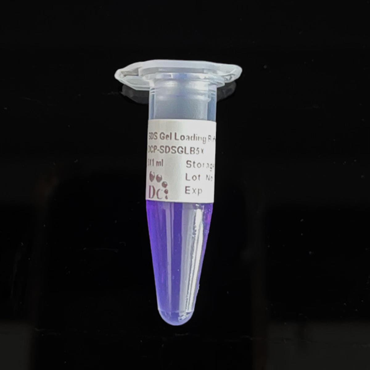
- Blue liquid appearance with integrated bromophenol blue tracking dye for easy gel-well loading
- Manufactured under ISO 13485-certified, CE-approved facilities
- Custom concentrations, additives, and pH available on request
- pH6.8
- Concentration5X
- AppearanceBlue liquid
- Filtration0.1 µm x2 + 0.04 µm x2
- SterilitySterile-filtered
- DNase ActivityNone detected
- RNase ActivityNone detected
- Storage-20°C
- Shelf Life1 year
- CustomizationAvailable on request
Engineered where standard loading buffers fall short
Conventional 0.22 µm-filtered loading buffers can carry subvisible particulates, inconsistent pH, and unverified nuclease activity into sensitive downstream molecular biology and microfluidic workflows. FluxMPS™ SDS Gel Loading Buffer [5X] is built to close those gaps.
Microchannel-safe purity
Sequential 0.1 µm and 0.04 µm membrane filtration removes particulates that could interfere with downstream SDS-PAGE and microfluidic sample-handling workflows.
Precise, consistent pH
Formulated at pH 6.8 with a defined Tris-HCl, SDS, glycerol, and TCEP system so every lot denatures and reduces protein samples the same way, run after run.
Ultrapure-grade water
Prepared with Ultrapure Type 1 water (18.2 MΩ·cm), reducing background contaminants in downstream gel and blot analysis.
Low background for imaging & assays
The integrated bromophenol blue tracking dye and ultra-clean formulation minimize background interference during electrophoresis, western blotting, and downstream imaging of MPS/OoC-derived protein samples.
Defined, traceable composition
Every lot is formulated with fixed concentrations of Tris-HCl, SDS, glycerol, TCEP, and bromophenol blue, and screened for DNase and RNase activity so protein sample prep stays reproducible.
Customization on demand
Concentration, pH, and additional chemicals, compounds, proteins, or supplements can be adjusted to match a specific sample-loading protocol — contact support@diagnocine.com.
Quadruple-stage filtration system
FluxMPS™ SDS Gel Loading Buffer [5X] is manufactured through a four-stage membrane filtration sequence — 0.1 µm membrane filtration twice followed by 0.04 µm membrane filtration twice — inside a sterile environment, engineered to keep particulate and bioburden out of sensitive downstream molecular biology and microfluidic sample-preparation workflows.
-
1
0.1 µm Pre-filtration I
First 0.1 µm membrane pass clears large particulates and aggregates from the Tris-HCl, SDS, glycerol, and TCEP formulation before downstream filtration.
-
2
0.04 µm Pre-filtration II
First 0.04 µm membrane pass retains fine particulates and bioburden ahead of final sterile filtration.
-
3
0.1 µm Sterile-filtration I
Second 0.1 µm membrane pass provides redundant particulate clearance immediately before final polishing.
-
4
0.04 µm Sterile-filtration II — Final Polish
Second 0.04 µm membrane pass delivers the final polish inside a sterile fill environment, helping prevent mycoplasma contamination — the smallest mycoplasma types measure about 0.2 microns.
Performance vs. conventional buffer
Sequential 0.1 µm and 0.04 µm filtration removes finer particulates than a single 0.22 µm pass, keeping SDS-PAGE sample buffer clean for downstream microfluidic and molecular biology workflows.

© Diagnocine® — DCP-SDSLB5X
Where DCP-SDSLB5X fits your workflow
From routine SDS-PAGE sample prep to protein analysis of organ-on-a-chip and microphysiological system cultures, FluxMPS™ SDS Gel Loading Buffer [5X] denatures, reduces, and tracks protein samples with ultra-clean, reproducible performance.
Automated Bioreactors & Robotics
An optional 0.01 µm (10 nm) ultra-filtered variant of FluxMPS™ SDS Gel Loading Buffer [5X] is available for automated bioreactor and robotic liquid-handling platforms where valve and sensor protection is critical.
- Total Particulate Exclusion: Removes particulates finer than the standard 0.04 µm polish for automated systems.
- Valve & Sensor Protection: Reduces the risk of microvalve and sensor fouling in automated dispensing lines.
- Extended Perfusion Stability: Supports consistent performance across long automated sample-preparation runs.
Inquiry Required: Contact support@diagnocine.com to request the 0.01 µm ultra-filtered variant of DCP-SDSLB5X for automated bioreactor and robotics platforms.
Micro Physiological System (MPS) & Chip
Prepares denatured, reduced protein lysates from OoC and MPS chip cultures for downstream SDS-PAGE and western blot analysis.
Wash, Dilution & Reconstitution
Denatures and reduces protein samples while adding density and a visible tracking dye for consistent gel-well loading.
iPSC-Derived Model Handling
Prepares iPSC-derived neuron, cardiomyocyte, and hepatocyte lysates for downstream protein expression analysis.
Endothelial & Primary Cell Perfusion
Supports protein analysis of endothelial and primary cell lysates recovered from perfusion-based vascular models.
ELISA, Blotting & Blocking
Loads denatured protein samples for western blot analysis following ELISA, IHC, or IF workflows.
Microscopy & Optical Sensing
Prepares protein samples from imaged live-cell and biosensor experiments for confirmatory SDS-PAGE analysis.
Measured and declared parameters
Every value below is sourced from the release specification and quality control record for DCP-SDSLB5X.
| Parameter | Specification |
|---|---|
| Formulation / Composition | Tris-HCl, SDS, Glycerol, TCEP, Bromophenol blue (5X concentrate) |
| Concentration | 5X |
| Appearance | Blue liquid |
| pH (USP <791>) | 6.8 |
| Format | 5 x 1 mL |
| Parameter | Specification |
|---|---|
| Sterility USP <71> | Filtered 0.1-micron membrane twice and 0.04-micron membrane twice in a sterile environment |
| DNase Activity | None detected (18 hr, room temperature, plasmid DNA challenge) |
| RNase Activity | None detected (18 hr, room temperature, ribosomal RNA challenge) |
| Water Quality USP <85> | Ultrapure Type 1 water (18.2 MΩ·cm) |
| Manufacturing Standard ISO 13485 | ISO 13485-certified, CE-approved facilities |
| Fill Environment | Sterile controlled environment |
| Parameter | Specification |
|---|---|
| Storage Temperature | -20°C |
| Shelf Life | 1 year |
| Parameter | Specification |
|---|---|
| Traceability | Assembled and quality-tested at DiagnoCine Precision, Totowa, New Jersey, USA |
| Manufacturing QMS ISO 13485 | ISO 13485-certified |
| Regulatory Alignment | CE-approved manufacturing facilities |
| Intended Use | Research Use Only (RUO) |
Full composition
DCP-SDSLB5X is a defined 5X denaturing loading buffer released on a per-lot basis against the concentrations below.
| Component | CAS Number | Concentration |
|---|---|---|
| Tris-HCl | 1185-53-1 | 250 mM |
| SDS | 151-21-3 | 10% SDS |
| Glycerol | 56-81-5 | 30% Glycerol |
| TCEP | 51805-45-9 | 50 mM |
| Bromophenol blue | 115-39-9 | 0.02% |
Manufacturing & compliance
DCP-SDSLB5X is manufactured, tested, and packaged under a controlled quality system.
ISO 13485:2016 QMS
Manufactured under ISO 13485-certified, CE-approved facilities (Suppliers of DiagnoCine Precision).
Ultrapure Type 1 Water
Formulated with Ultrapure Type 1 water (18.2 MΩ·cm) consistent with USP <85>.
Sterile Fill & Finish
Filtered and packaged in a sterile controlled environment to prevent mycoplasma contamination.
Micro-Batch Precision
All final packaging, quality assurance, and testing are completed at the DiagnoCine R&D and Quality Testing Center; customization and assembly occur at DiagnoCine Precision in Totowa, New Jersey, USA.
Sterile Filtration
0.1-micron membrane filtration twice and 0.04-micron membrane filtration twice.
DNase & RNase Screening
None detected after 18-hour challenge testing at room temperature.
pH Verification USP <791>
Released at pH 6.8.
Documentation / CoA
Certificate of Analysis available on request.
How DCP-SDSLB5X compares
A qualitative comparison against typical conventional loading buffer manufacturing.
| Parameter | DCP-SDSLB5X (FluxMPS™) | Conventional loading buffer (0.22 µm filtered) | Standard alternative (0.22 µm filtered) |
|---|---|---|---|
| Denaturing/reducing formulation (Tris-HCl, SDS, Glycerol, TCEP, BPB) | check_circle | cancel | cancel |
| Final filtration pore size | 0.04 µm | 0.22 µm | 0.22 µm |
| Number of filtration stages | 4 | 1 | 1 |
| DNase & RNase screening | check_circle | cancel | cancel |
| Sterility verification | check_circle | cancel | cancel |
| Water quality (Type 1, 18.2 MΩ·cm) | check_circle | cancel | cancel |
| Manufacturing QMS (ISO 13485) | check_circle | cancel | cancel |
| Microfluidic / OoC channel compatibility | check_circle | cancel | cancel |
| pH consistency (lot-to-lot) | check_circle | cancel | check_circle |
| Custom formulation available | check_circle | cancel | cancel |
Frequently asked questions
Answers to common questions about DCP-SDSLB5X.
Supporting literature
Curated peer-reviewed literature relevant to SDS-PAGE sample preparation and organ-on-a-chip protein analysis.
- Laemmli UK. Cleavage of structural proteins during the assembly of the head of bacteriophage T4. Nature. 1970. doi:10.1038/227680a0
- Shapiro AL, Vinuela E, Maizel JV. Molecular weight estimation of polypeptide chains by electrophoresis in SDS-polyacrylamide gels. Biochem Biophys Res Commun. 1967. doi:10.1016/S0006-291X(67)80055-X
- Rath A, Glibowicka M, Nadeau VG, Chen G, Deber CM. Detergent binding explains anomalous SDS-PAGE migration of membrane proteins. Proc Natl Acad Sci USA. 2009. doi:10.1073/pnas.0813167106
- Getz EB, Xiao M, Chakrabarty T, Cooke R, Selvin PR. A comparison between the sulfhydryl reductants tris(2-carboxyethyl)phosphine and dithiothreitol for use in protein biochemistry. Anal Biochem. 1999. doi:10.1006/abio.1999.4203
- Chrambach A, Rodbard D. Polyacrylamide gel electrophoresis. Science. 1971. doi:10.1126/science.172.3982.440
- Bhatia SN, Ingber DE. Microfluidic organs-on-chips. Nat Biotechnol. 2014. doi:10.1038/nbt.2989
- Huh D, Matthews BD, Mammoto A, Montoya-Zavala M, Hsin HY, Ingber DE. Reconstituting organ-level lung functions on a chip. Science. 2010. doi:10.1126/science.1188302
- Zhang B, Korolj A, Lai BFL, Radisic M. Advances in organ-on-a-chip engineering. Nat Rev Mater. 2018. doi:10.1038/s41578-018-0034-7
- Low LA, Mummery C, Berridge BR, Austin CP, Tagle DA. Organs-on-chips: into the next decade. Nat Rev Drug Discov. 2021. doi:10.1038/s41573-020-0079-3