

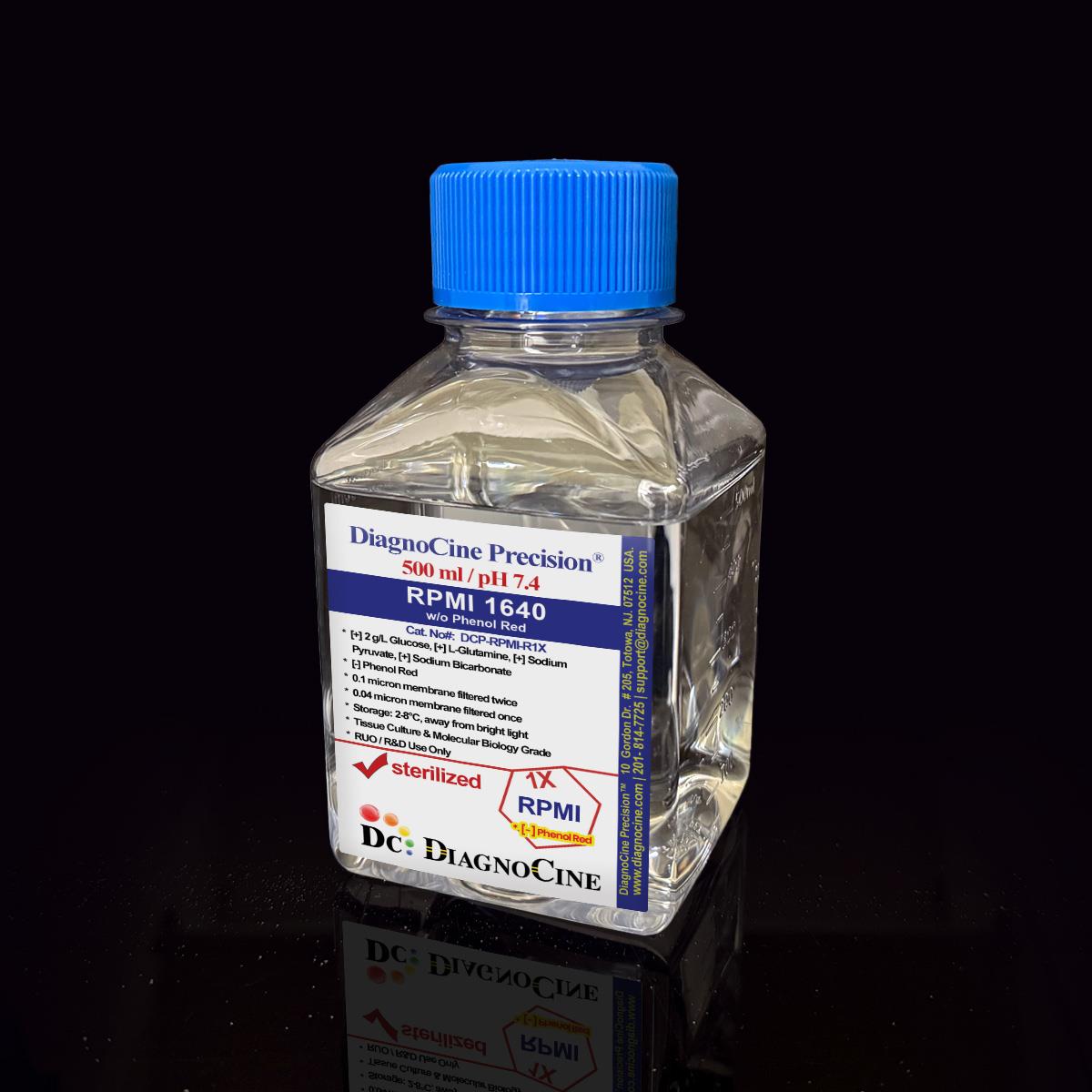
FluxMPS™ RPMI 1640 Medium w/o Phenol Red: 1X Liquid
FluxMPS™ RPMI 1640 Medium w/o Phenol Red
FluxMPS™ DCP-RPMI-R1X is a Microfluidics Suitable, quadruple-stage ultra-filtered (0.1 µm ×2 + 0.04 µm ×2) RPMI 1640 formulation without phenol red, engineered for lymphocyte, hematopoietic and immune cell models on organ-on-a-chip (OoC) and microphysiological system (MPS) platforms. A quadruple-stage train (0.1 µm ×2 + 0.04 µm ×2) reaches a 0.04 µm final cut-off, five times finer than the 0.22 µm membranes used for conventional sterile filtration.
- Glucose source: 2000 mg/L (2.0 g/L) D-glucose for standard RPMI energy metabolism
- L-Glutamine 300 mg/L and Sodium Pyruvate 110 mg/L support lymphocyte and hematopoietic cell energy demands
- Glutathione (reduced, 1.0 mg/L) included — antioxidant support for redox-sensitive immune cell types
- Quadruple-stage filtration (0.1 µm ×2 + 0.04 µm ×2) reaching a 0.04 µm final pore size for sub-mycoplasma-range purity
- Endotoxin release specification: < 0.05 EU/mL (LAL, USP <85>) — relevant to TLR4-sensitive immune cell assays
- Phenol red excluded — eliminates 520–560 nm autofluorescence for flow cytometry, confocal imaging and hormone-sensitive assays
- Manufactured under an ISO 13485:2016 quality management system; final QC at Diagnocine, Totowa NJ
- Custom pH, nutrient concentration and HEPES buffering available on request
- Media familyRPMI 1640
- Glucose2000 mg/L (2.0 g/L)
- L-Glutamine300 mg/L
- Sodium Pyruvate110 mg/L
- pH (USP <791>)7.4
- Osmolality (USP <785>)280–320 mOsm/kg H2O
- Endotoxin (USP <85>)< 0.05 EU/mL
- Filtration0.1 µm ×2 + 0.04 µm ×2 (Quadruple-stage)
- Storage2–8°C, protect from light
- Shelf Life12 months from date of manufacture, unopened
Engineered where standard media fails
Conventional 0.22 µm–filtered RPMI passes mycoplasma-scale particles, subvisible particulates, and phenol red autofluorescence that confound immune cell assays. FluxMPS™ is built to remove these failure modes at the filtration and formulation level.
Microchannel-safe purity
0.04 µm final filtration; USP <788> Method 1 (light obscuration) particulate compliance. Particle-controlled media for suspension immune cell OoC and flow cytometry.
Immune cell–optimized formulation
RPMI 1640 contains reduced glutathione and a balanced amino acid / vitamin profile suited to lymphocyte and hematopoietic cell culture.
Purity-controlled water
Manufactured with trace-metal and total organic carbon (TOC)-controlled process water as part of the ISO 13485:2016 quality system.
Low background for imaging
Phenol red excluded from this formulation, reducing baseline particulate and optical interference for confocal and PE-channel flow cytometry work.
Rich nutrient profile
RPMI's specialized amino acid and vitamin profile is delivered through a 0.04 µm final-filtered process for OoC use.
Customization on demand
pH, nutrient concentrations, HEPES, and component modifications available. Contact support@diagnocine.com.
Quadruple-stage filtration system
Four serial filtration passes — two dedicated prefilter + final-filter pairs — reaching a final 0.04 µm polish.
-
1
0.1 µm Prefiltration I
Removes large particulate, cell debris and protein aggregates; protects the first 0.04 µm cartridge.
-
2
0.04 µm Final filtration I
First 0.04 µm pass; retains sub-micron particulates and microaggregates that pass a standard 0.22 µm filter.
-
3
0.1 µm Prefiltration II
Second dedicated prefilter, protecting the second 0.04 µm cartridge from particulate load.
-
4
0.04 µm Final filtration II — Polish
Ultimate polishing filter prior to aseptic fill & finish.
Performance vs. conventional media

© Diagnocine® — DCP-RPMI-R1X
Immune cell OoC and hematopoietic applications
FluxMPS™ DCP-RPMI-R1X — RPMI 1640 w/o phenol red — delivers 0.04 µm filtered, Microfluidics Suitable purity for lymphocyte, hematopoietic and cancer cell OoC applications.
Automated Bioreactors & Robotics
An optional 0.01 µm (10 nm) ultra nano-filtered MPS Grade variant of this formulation is available on request for automated perfusion and robotic handling systems.
- Total Particulate Exclusion: 0.01 µm filtration removes nanoparticulate aggregates
- Valve & Sensor Protection: Reduces micro-fouling risk in automated perfusion systems
- Extended Perfusion Stability: Consistent nutrient delivery over long-duration culture
Inquiry Required: Contact support@diagnocine.com for the 0.01 µm MPS Grade variant.
T Cell & Lymphocyte Culture
RPMI 1640 is the standard for primary T cells, B cells, NK cells, and monocytes. 0.04 µm filtration reduces particulate load relevant to immune assay signal.
Leukemia & Lymphoma Lines
RPMI supports NCI-60 cancer lines, Jurkat, Raji, K562, HL-60, and hematopoietic cancer lines where DMEM would alter proliferation and signaling.
Immune Cell OoC
0.04 µm filtered RPMI for tumor-immune interaction chips, vascular-immune OoC, and lymph node-on-chip microphysiological systems (MPS).
CAR-T & TIL Expansion
Endotoxin release specification < 0.05 EU/mL is relevant to CAR-T manufacturing and TIL expansion protocols sensitive to TLR4 activation.
Immune Cell Metabolic Flux
Defined RPMI base for glycolysis stress tests and ¹³C isotope tracing of lymphocyte activation states. Not compatible with Agilent Seahorse XF assays, which require bicarbonate-free, phenol red-free medium.
Flow Cytometry & Confocal
Phenol red–free formulation reduces autofluorescence for PE-channel flow cytometry and immune cell confocal imaging.
Analytical release specifications
Every lot released against the full specification matrix. Pack sizes: 500 mL, 1000 mL. CoA: support@diagnocine.com.
| Parameter | Specification |
|---|---|
| Formulation | [+] L-Glutamine, [+] Sodium Bicarbonate, [+] Calcium, [+] Magnesium, [+] 2 g/L Glucose, [+] Sodium Pyruvate | [-] Phenol Red |
| Appearance | Colorless to pale yellow, clear solution |
| Glucose | 2000 mg/L (2.0 g/L) |
| pH USP <791> | 7.4 |
| Osmolality USP <785> | 280–320 mOsm/kg H2O |
| Total ingredients | 40 |
| Parameter | Specification |
|---|---|
| Endotoxin USP <85> BET | < 0.05 EU/mL |
| Sterility USP <71> | No growth / 14 days |
| Mycoplasma | 0.1 µm mycoplasma-retentive filtration (not tested per lot) |
| Particulate ≥10 µm USP <788> Method 1 | NMT 25/mL |
| Particulate ≥25 µm USP <788> Method 1 | NMT 3/mL |
| Manufacturing std. | ISO 13485:2016 |
| Fill environment | ISO Class 5 (Class 100) |
| Parameter | Specification |
|---|---|
| Storage temperature | 2–8°C, away from light |
| Freeze-thaw | Do not freeze |
| Shelf life | 12 months from date of manufacture, unopened |
| Shipping condition | Cold pack |
| CO2 requirement | 5% CO2 required (standard RPMI 1640 bicarbonate buffering system) |
| Parameter | Specification |
|---|---|
| Raw material grade | Reagent / cell culture grade |
| Traceability | Full lot traceability per ISO 13485 |
| Manufacturing QMS ISO | ISO 13485:2016 certified |
| UNSPSC | 41116155 — Molecular biology and cell culture growth media (UNv260801) |
| Regulatory alignment | 21 CFR Part 820 (QMSR) aligned |
| Production method | Micro-batch, per-lot QC release |
| Intended use | Research Use Only (RUO) |
Full composition (mg/L)
RPMI 1640: 40 ingredients verified per lot with CAS numbers for raw-material traceability. Contains reduced glutathione and a balanced amino acid / vitamin profile suited to lymphocyte and hematopoietic cell culture.
| Component | CAS Number | mg/L |
|---|---|---|
| INORGANIC SALTS | ||
| Calcium nitrate tetrahydrate | 13477-34-4 | 100.000 |
| Magnesium sulfate anhydrous | 7487-88-9 | 48.840 |
| Potassium chloride | 7447-40-7 | 400.000 |
| Sodium bicarbonate | 144-55-8 | 2000.000 |
| Sodium chloride | 7647-14-5 | 6000.00 |
| Sodium phosphate dibasic anhydrous | 7558-79-4 | 800.000 |
| Component | CAS Number | mg/L |
|---|---|---|
| AMINO ACIDS | ||
| Glycine | 56-40-6 | 10.000 |
| L-Arginine hydrochloride | 1119-34-2 | 241.000 |
| L-Asparagine | 70-47-3 | 50.000 |
| L-Aspartic acid | 56-84-8 | 20.000 |
| L-Cystine dihydrochloride | 30925-07-6 | 65.200 |
| L-Glutamic acid | 56-86-0 | 20.000 |
| L-Glutamine | 56-85-9 | 300.000 |
| L-Histidine hydrochloride monohydrate | 5934-29-2 | 20.960 |
| L-Hydroxyproline | 51-35-4 | 20.000 |
| L-Isoleucine | 73-32-5 | 50.000 |
| L-Leucine | 61-90-5 | 50.000 |
| L-Lysine hydrochloride | 657-27-2 | 40.000 |
| L-Methionine | 63-68-3 | 15.000 |
| L-Phenylalanine | 63-91-2 | 15.000 |
| L-Proline | 147-85-3 | 20.000 |
| L-Serine | 56-45-1 | 30.000 |
| L-Threonine | 72-19-5 | 20.000 |
| L-Tryptophan | 73-22-3 | 5.000 |
| L-Tyrosine Disodium Salt | 69847-45-6 | 28.830 |
| L-Valine | 72-18-4 | 20.000 |
| Component | CAS Number | mg/L |
|---|---|---|
| VITAMINS | ||
| Choline chloride | 67-48-1 | 3.000 |
| D-Biotin | 58-85-5 | 0.200 |
| D-Ca-Pantothenate | 137-08-6 | 0.250 |
| Folic acid | 59-30-3 | 1.000 |
| Niacinamide | 98-92-0 | 1.000 |
| Pyridoxine hydrochloride | 58-56-0 | 1.000 |
| Riboflavin | 83-88-5 | 0.200 |
| Thiamine hydrochloride | 67-03-8 | 1.000 |
| Vitamin B12 | 68-19-9 | 0.005 |
| i-Inositol | 87-89-8 | 35.000 |
| p-Amino benzoic acid (PABA) | 150-13-0 | 1.000 |
| OTHERS | ||
| D-Glucose | 50-99-7 | 2000.000 |
| Glutathione reduced | 70-18-8 | 1.000 |
| Sodium Pyruvate | 113-24-6 | 110.000 |
Manufacturing & compliance
Every FluxMPS™ product manufactured and released under a rigorous multi-layer quality system.
ISO 13485:2016 Quality Management
Manufactured under an ISO 13485:2016-certified quality management system. Final QC at Diagnocine R&D Center, Totowa, NJ, USA.
Quadruple-Stage Filtration
0.1 µm ×2 + 0.04 µm ×2 filtration train applied to every batch.
ISO Class 5 Fill & Finish
Aseptic fill in validated ISO Class 5 (Class 100) laminar-flow workstations.
Micro-Batch Precision
Small-batch, per-lot tested — Certificate of Analysis available for every lot.
Endotoxin — USP <85> BET
LAL assay; release specification < 0.05 EU/mL per batch.
Particulate — USP <788> Method 1
Light obscuration: NMT 25/mL (≥10 µm), NMT 3/mL (≥25 µm).
Osmolality — USP <785>
Target: 280–320 mOsm/kg H2O.
Documentation & CoA
Full CoA with raw-material traceability available on request.
- Endotoxin — LAL assay, USP <85> Bacterial Endotoxins Test; assay sensitivity 0.005 EU/mL; release specification < 0.05 EU/mL
- pH, osmolality, conductivity, appearance and clarity
- Sterility
How DCP-RPMI-R1X compares
FluxMPS™ DCP-RPMI-R1X vs. conventional 0.22 µm–filtered RPMI formulations.
| Parameter | DCP-RPMI-R1X (FluxMPS™) | Conventional RPMI 1640 (0.22 µm filtered) |
Standard DMEM/RPMI (0.22 µm filtered) |
|---|---|---|---|
| Grade | Microfluidics Suitable | Not designated | Not designated |
| RPMI 1640 without Phenol Red — autofluorescence-reduced for flow cytometry, confocal, and hormone-sensitive assays | check_circle Yes | cancel No | cancel No |
| Final filtration pore size | 0.04 µm | 0.22 µm | 0.22 µm |
| Number of filtration stages | 4 (Quadruple) | 1 | 1 |
| Mycoplasma barrier filtration | check_circle Yes (0.1 µm) | cancel No | cancel No |
| Endotoxin (release specification) | FluxMPS™ — < 0.05 EU/mL | Corning classical liquid media — < 0.25 EU/mL Sigma-Aldrich DMEM complete medium — ≤ 2 EU/mL Gibco classical DMEM — Not specified (recorded per lot) |
|
| USP <788> particulate tested | check_circle Yes (Method 1) | cancel Not specified | cancel Not specified |
| Manufacturing QMS | ISO 13485:2016 | Not specified | Not specified |
| Microfluidic channel compatibility | check_circle Microfluidics Suitable (0.04 µm) | cancel Risk of clogging | cancel Risk of clogging |
| Custom formulation available | check_circle Yes | cancel No | cancel No |
Comparison figures from published supplier specifications, accessed 2026-09-02. Suppliers that publish no numeric endotoxin specification are shown as "Not specified".
Frequently asked questions
Common questions about FluxMPS™ DCP-RPMI-R1X — RPMI 1640 w/o Phenol Red.
Supporting literature
Key publications supporting RPMI 1640 in immune cell culture and OoC applications.
- Moore GE, et al. Culture of normal human leukocytes. JAMA. 1967;199:519–524. doi:10.1083/jcb.1.3.273
- Huh D, et al. Reconstituting organ-level lung functions on a chip. Science. 2010;328:1662–1668. doi:10.1126/science.1188302
- Bhatia SN, Ingber DE. Microfluidic organs-on-chips. Nat Biotechnol. 2014;32:760–772. doi:10.1038/nbt.2989
- Novak R, et al. Robotic fluidic coupling and interrogation of multiple vascularized organ chips. Nat Biomed Eng. 2020;4:407–420. doi:10.1038/s41551-019-0497-x
- Jang KJ, et al. Human kidney proximal tubule-on-a-chip. Integr Biol. 2013;5:1119–1129. doi:10.1039/c3ib40049b
- Schimek K, et al. Integrating biological vasculature into a multi-organ-chip microsystem. Lab Chip. 2013;13:3588–3598. doi:10.1039/c3lc50217a
- Luni C, et al. High-efficiency cellular reprogramming with microfluidics. Nat Methods. 2016;13:446–452. doi:10.1038/nmeth.3832
- Sung JH, et al. Microfabricated mammalian organ systems. Lab Chip. 2013;13:1201–1212. doi:10.1039/c3lc41017j
- Gehring MA, et al. Immune-cell microphysiological systems for lymphocyte biology. Trends Biotechnol. 2019;37:591–603. doi:10.1016/j.tibtech.2018.11.001