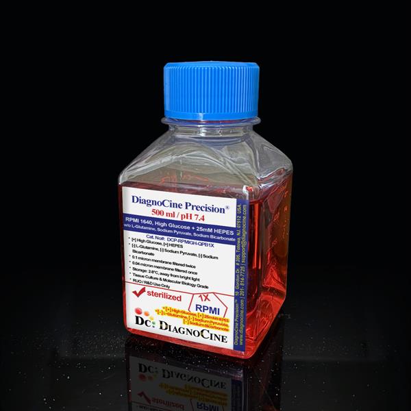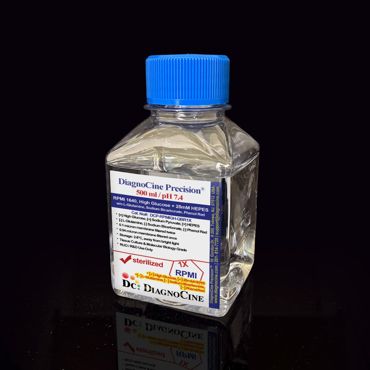
FluxMPS™ RPMI 1640, High Glucose + 25mM HEPES w/o Glutamine, Bicarbonate, Phenol Red
FluxMPS™ RPMI 1640, High Glucose + 25mM HEPES w/o Glutamine, Bicarbonate, Phenol Red
A Microfluidics Suitable, ready-to-use 1× RPMI 1640 medium with 4500 mg/L D-glucose, 25 mM HEPES and sodium pyruvate — supplied without L-glutamine, sodium bicarbonate or phenol red. Quadruple-stage filtration (0.1 µm membrane ×2, 0.04 µm membrane ×2) delivers a low-particulate fluid engineered for microfluidic channels, organ-on-a-chip devices and demanding suspension and adherent cultures.
- High glucose (4.5 g/L) RPMI 1640 with 25 mM HEPES buffering; formulated without L-glutamine, sodium bicarbonate or phenol red
- Quadruple-stage filtration: 0.1 µm membrane ×2 + 0.04 µm membrane ×2, engineered for microchannel-safe fluidics
- Endotoxin release specification: less than 0.05 EU/mL (LAL, USP <85>)
- Sodium pyruvate (110 mg/L) included as an alternate carbon/energy substrate alongside high glucose
- L-glutamine-free design supports user-defined glutamine or GlutaMAX™ supplementation and improved shelf stability
- 25 mM HEPES buffering supports pH stability in closed and limited-CO₂ microfluidic and perfusion systems
- Manufactured under an ISO 13485:2016 quality management system; final QC and packaging at Diagnocine, Totowa, NJ
- pH, glucose, salts, HEPES molarity and nutrient composition customizable on request
- Glucose4500 mg/L (4.5 g/L)
- L-GlutamineNone (add on use)
- HEPES5958 mg/L (25 mM)
- Sodium Pyruvate110 mg/L
- pH (USP <791>)7.4
- Osmolality (mOsm/kg)230–270
- Endotoxin (LAL, USP <85>)< 0.05 EU/mL
- Filtration0.1 µm ×2 + 0.04 µm ×2
- Storage2–8°C, protect from light
- Shelf Life12 months from date of manufacture, unopened
Engineered where standard media reach their limits
Conventional 0.22 µm-filtered media can still carry sub-visible particulates and the smallest mycoplasma forms (approximately 0.2–0.3 µm). In microfluidic channels and organ-on-a-chip devices, such particulates accumulate at junctions, foul sensors and corrupt optical readouts. FluxMPS™ RPMI 1640 finishes with a 0.04 µm final filtration pass, retaining particulates and mycoplasma-scale bodies that a single 0.22 µm filter leaves behind[8].
Microchannel-safe purity
A 0.04 µm final filter retains fine particulates and mycoplasma-scale bodies, protecting narrow microfluidic channels from occlusion.
Total metabolic control
Bicarbonate-free, L-glutamine-free design lets you define the carbon and nitrogen source — suited to Warburg effect and metabolic flux studies[3].
Buffered for open and closed systems
25 mM HEPES supplements the medium's buffering so pH stays stable in limited-CO₂ and closed perfusion setups[4].
Low background for imaging
Phenol-red-free formulation reduces optical background from media color, and low-particulate filtration supports clean fields for confocal and biosensor work. Note that riboflavin (a required B-vitamin, 0.2 mg/L) is intrinsically fluorescent; account for it when designing fluorescence-based assays.
Rich, defined nutrient profile
A full RPMI 1640 amino acid and vitamin spectrum plus sodium pyruvate supports lymphocytes, hybridomas and carcinoma lines[1].
Customization on demand
pH, glucose, salts, HEPES and supplements adjustable per request — contact support@diagnocine.com.
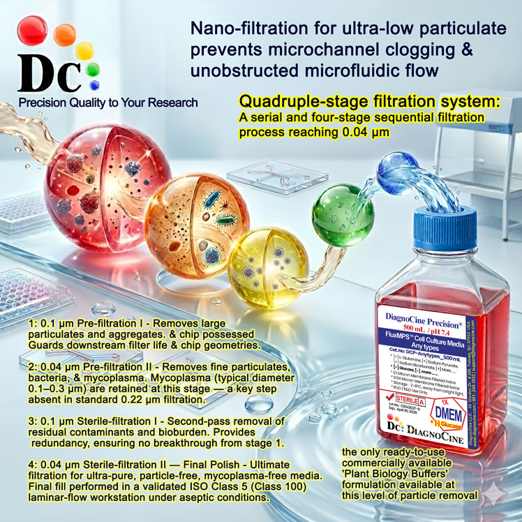
Quadruple-stage filtration system
Every lot is filtered through a paired prefilter-plus-final-filter train, run twice in series — 0.1 µm, 0.04 µm, 0.1 µm, 0.04 µm — rather than a single 0.22 µm pass. Each 0.04 µm final filter is protected by its own dedicated 0.1 µm prefilter; the second pair provides full redundancy. Because the smallest mycoplasma forms measure roughly 0.2–0.3 µm, the 0.04 µm final membranes are well below that size.
-
01
0.1 µm Prefiltration I
First 0.1 µm membrane pass removes large particulates and aggregates, protecting the first 0.04 µm cartridge.
-
02
0.04 µm Final filtration I
First 0.04 µm membrane pass retains fine particulates and mycoplasma-scale bodies that a 0.22 µm filter would pass.
-
03
0.1 µm Prefiltration II
Second, dedicated 0.1 µm membrane pass protects the second 0.04 µm cartridge and further reduces fine particulate burden.
-
04
0.04 µm Final filtration II — Polish
Second 0.04 µm membrane delivers the ultimate polishing pass ahead of aseptic fill.
Performance vs. single 0.22 µm filtration
Two 0.1 µm + 0.04 µm filter pairs, run in series, retain particulates and mycoplasma-scale bodies that pass through a single 0.22 µm filter, lowering the subvisible particulate load delivered into microfluidic channels.
© Diagnocine® — DCP-RPMIGH-QBR1X
From microphysiological systems to classic culture
The bicarbonate-free, HEPES-buffered, high-glucose formulation suits both microphysiological platforms and the lymphocyte, hybridoma and carcinoma cultures RPMI 1640 was designed for[1].
Automated Bioreactors & Robotics
For automated perfusion bioreactors and liquid-handling robotics, an optional MPS Grade 0.01 µm (10 nm) ultra-filtered variant of this medium can be produced on request to further reduce subvisible particulates that challenge fine valves and inline sensors.
- Total particulate exclusion for narrow fluidic paths
- Valve & sensor protection in unattended runs
- Extended perfusion stability between feeds
Inquiry Required: the MPS Grade 0.01 µm (10 nm) variant is made to order — contact support@diagnocine.com.
Microphysiological System (MPS) & Chip
Warburg Effect & Metabolic Research
Lymphocyte & Hybridoma Culture
Leukemia & Bone Marrow Models
Defined-Carbon Metabolic Flux
Phenol-Red-Free Microscopy
Technical specifications
All values reflect the product Certificate of Analysis basis at 1× concentration.
| Parameter | Specification |
|---|---|
| Formulation | RPMI 1640, high glucose; contains HEPES, calcium, magnesium, glucose, sodium pyruvate; without L-glutamine, sodium bicarbonate, phenol red |
| Appearance | Pale yellow, clear solution |
| pH (USP <791>) | 7.4 |
| Osmolality (USP <785>) | 230–270 mOsm/kg H2O USP |
| D-Glucose | 4500 mg/L |
| L-Glutamine | Not added |
| Sodium Pyruvate | 110 mg/L |
| HEPES | 5958 mg/L (25 mM) |
| Phenol Red | None |
| Parameter | Specification |
|---|---|
| Endotoxin | < 0.05 EU/mL (LAL, USP <85>) BET |
| Sterility | No growth, 14 days, per USP <71> USP |
| Mycoplasma | 0.1 µm and 0.04 µm mycoplasma-retentive filtration (not tested per lot) |
| Filtration | 0.1 µm ×2 + 0.04 µm ×2 |
| Cultural response | Growth-promotion verified vs. control medium |
| Manufacturing std. | ISO 13485:2016-certified facilities ISO |
| Regulatory | ISO 13485:2016 aligned; Research Use Only (RUO) |
| Parameter | Specification |
|---|---|
| Storage temperature | 2–8°C, protect from light |
| Shelf life | 12 months from date of manufacture, unopened |
| Use by | Expiry date on product label |
| Shipping condition | Ambient / cold pack |
| CO₂ requirement | HEPES-buffered; reduced CO₂ dependence (validate per cell line) |
| Parameter | Specification |
|---|---|
| Raw material grade | Cell-culture grade |
| Traceability | Lot-controlled; CoA per lot |
| Manufacturing QMS | ISO 13485:2016 ISO |
| UNSPSC | 41116155 — Molecular biology and cell culture growth media (UNv260801) |
| Regulatory alignment | ISO 13485:2016 aligned; Research Use Only (RUO) |
| Final QC / packaging | Diagnocine R&D and Quality Testing Center, Totowa NJ |
| Intended use | Research Use Only (RUO) |
Full composition (mg/L)
Complete RPMI 1640 high-glucose formulation as released per lot. Total: 39 components. Ingredient names and mg/L values are reproduced from the product specification; CAS numbers are added for reference where established.
| Component | CAS Number | mg/L |
|---|---|---|
| INORGANIC SALTS | ||
| Calcium nitrate tetrahydrate | 13477-34-4 | 100.000 |
| Magnesium sulfate anhydrous | 7487-88-9 | 48.840 |
| Potassium chloride | 7447-40-7 | 400.000 |
| Sodium chloride | 7647-14-5 | 6000.000 |
| Sodium phosphate dibasic anhydrous | 7558-79-4 | 800.000 |
| Component | CAS Number | mg/L |
|---|---|---|
| AMINO ACIDS | ||
| Glycine | 56-40-6 | 10.000 |
| L-Arginine hydrochloride | 1119-34-2 | 241.000 |
| L-Asparagine | 70-47-3 | 50.000 |
| L-Aspartic acid | 56-84-8 | 20.000 |
| L-Cystine dihydrochloride | 30925-07-6 | 65.200 |
| L-Glutamic acid | 56-86-0 | 20.000 |
| L-Histidine hydrochloride monohydrate | 5934-29-2 | 20.960 |
| L-Hydroxyproline | 51-35-4 | 20.000 |
| L-Isoleucine | 73-32-5 | 50.000 |
| L-Leucine | 61-90-5 | 50.000 |
| L-Lysine hydrochloride | 657-27-2 | 40.000 |
| L-Methionine | 63-68-3 | 15.000 |
| L-Phenylalanine | 63-91-2 | 15.000 |
| L-Proline | 147-85-3 | 20.000 |
| L-Serine | 56-45-1 | 30.000 |
| L-Threonine | 72-19-5 | 20.000 |
| L-Tryptophan | 73-22-3 | 5.000 |
| L-Tyrosine Disodium Salt | — | 28.830 |
| L-Valine | 72-18-4 | 20.000 |
| Component | CAS Number | mg/L |
|---|---|---|
| VITAMINS | ||
| Choline chloride | 67-48-1 | 3.000 |
| D-Biotin | 58-85-5 | 0.200 |
| D-Ca-Pantothenate | 137-08-6 | 0.250 |
| Folic acid | 59-30-3 | 1.000 |
| Niacinamide | 98-92-0 | 1.00 |
| Pyridoxine hydrochloride | 58-56-0 | 1.00 |
| Riboflavin | 83-88-5 | 0.200 |
| Thiamine hydrochloride | 67-03-8 | 1.000 |
| Vitamin B12 | 68-19-9 | 0.005 |
| i-Inositol | 87-89-8 | 35.000 |
| p-Amino benzoic acid (PABA) | 150-13-0 | 1.000 |
| OTHERS | ||
| D-Glucose | 50-99-7 | 4500.000 |
| Glutathione reduced | 70-18-8 | 1.000 |
| HEPES | 7365-45-9 | 5958.000 |
| Sodium pyruvate | 113-24-6 | 110.000 |
Manufacturing & compliance
Produced under an ISO 13485-certified quality management system, with final packaging, quality assurance and testing at the Diagnocine R&D and Quality Testing Center, Totowa, New Jersey, USA.
ISO 13485:2016 QMS
Manufactured under a controlled quality management system certified to ISO 13485:2016.
Quadruple-stage filtration
0.1 µm (×2) + 0.04 µm (×2) membrane train for low-particulate fluid.
Per-lot QC & CoA
Appearance, pH, osmolality, sterility, endotoxin and cultural response verified and documented per lot.
Finished in Totowa, NJ
Final QC, packaging and customization performed at Diagnocine, Totowa, New Jersey, USA.
Endotoxin — USP <85>
Bacterial endotoxin release specification: less than 0.05 EU/mL, LAL assay, assay sensitivity 0.005 EU/mL.
Sterility — USP <71>
No bacterial or fungal growth after 14 days incubation per USP specification.
Osmolality — USP <785>
230–270 mOsm/kg H2O at 1× concentration.
Documentation / CoA
Certificate of Analysis available per lot on request.
- Endotoxin — LAL assay, USP <85> Bacterial Endotoxins Test; assay sensitivity 0.005 EU/mL; release specification < 0.05 EU/mL
- pH, osmolality, conductivity, appearance and clarity
- Sterility
How DCP-RPMIGH-QBR1X compares
Versus conventional single-pass 0.22 µm-filtered RPMI 1640 formulations, and against published supplier endotoxin specifications.
| Parameter | DCP-RPMIGH-QBR1X (FluxMPS™) | Conventional RPMI 1640 (0.22 µm) | Standard HEPES RPMI (0.22 µm) |
|---|---|---|---|
| Grade | Microfluidics Suitable | Not specified | Not specified |
| Formulation | High glucose, 25 mM HEPES, w/o L-glutamine / bicarbonate / phenol red | High glucose, varies | With HEPES, varies |
| Final filtration pore size | 0.04 µm | 0.22 µm | 0.22 µm |
| Number of filtration passes | 4 (0.1 µm ×2 + 0.04 µm ×2) | 1 | 1 |
| Mycoplasma-retentive filtration | check_circle | cancel | cancel |
| Endotoxin (release specification) | FluxMPS™ — < 0.05 EU/mL | Corning classical liquid media — < 0.25 EU/mL Sigma-Aldrich DMEM complete medium — ≤ 2 EU/mL Gibco classical DMEM — Not specified (recorded per lot) |
|
| Phenol-red-free (low background) | check_circle | cancel | Varies |
| Microfluidic channel compatibility | check_circle | Limited | Limited |
| Manufacturing QMS | ISO 13485:2016 | Varies | Varies |
| Custom formulation | check_circle | cancel | Varies |
Comparison figures from published supplier specifications, accessed 2 September 2026. Suppliers that publish no numeric endotoxin specification are shown as "Not specified".
Available pack sizes: 500 mL, 1000 mL.
Frequently asked questions
Common questions about formulation, filtration and handling.
Supporting literature
Peer-reviewed literature relevant to RPMI 1640, HEPES buffering, organ-on-a-chip culture and mycoplasma control.
- Moore GE, Gerner RE, Franklin HA. Culture of normal human leukocytes. JAMA. 1967;199(8):519-524. doi:10.1001/jama.1967.03120080053007
- Bhatia SN, Ingber DE. Microfluidic organs-on-chips. Nat Biotechnol. 2014;32(8):760-772. doi:10.1038/nbt.2989
- Vander Heiden MG, Cantley LC, Thompson CB. Understanding the Warburg effect: the metabolic requirements of cell proliferation. Science. 2009;324(5930):1029-1033. doi:10.1126/science.1160809
- Good NE, Winget GD, Winter W, et al. Hydrogen ion buffers for biological research. Biochemistry. 1966;5(2):467-477. doi:10.1021/bi00866a011
- Esch EW, Bahinski A, Huh D. Organs-on-chips at the frontiers of drug discovery. Nat Rev Drug Discov. 2015;14(4):248-260. doi:10.1038/nrd4539
- Huh D, Matthews BD, Mammoto A, et al. Reconstituting organ-level lung functions on a chip. Science. 2010;328(5986):1662-1668. doi:10.1126/science.1188302
- Liberti MV, Locasale JW. The Warburg effect: how does it benefit cancer cells? Trends Biochem Sci. 2016;41(3):211-218. doi:10.1016/j.tibs.2015.12.001
- Drexler HG, Uphoff CC. Mycoplasma contamination of cell cultures: incidence, sources, effects, detection, elimination, prevention. Cytotechnology. 2002;39(2):75-90. doi:10.1023/A:1022913015916
- Eagle H. Amino acid metabolism in mammalian cell cultures. Science. 1959;130(3373):432-437. doi:10.1126/science.130.3373.432
- Zhang B, Korolj A, Lai BFL, Radisic M. Advances in organ-on-a-chip engineering. Nat Rev Mater. 2018;3:257-278. doi:10.1038/s41578-018-0034-7