RIPA Buffer [1X]
FluxMPS™ RIPA Buffer [1X]
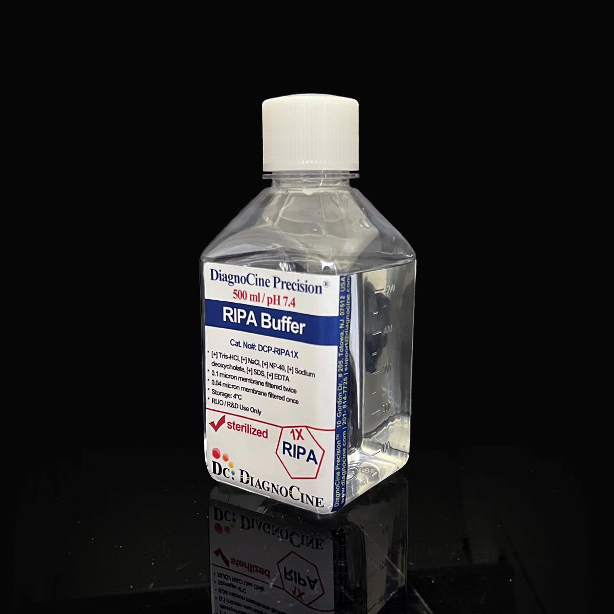
A sterile, ultrapure radioimmunoprecipitation assay (RIPA) lysis buffer formulated at pH 7.4 with Tris-HCl, NaCl, Nonidet P-40, sodium deoxycholate, SDS, and EDTA. Filtered 0.1-micron membrane twice and 0.04-micron membrane twice for microchannel-safe purity, this buffer extracts cytoplasmic, membrane, and nuclear proteins from adherent and suspension-cultured cells alike.
- Sterile, ultrapure RIPA lysis buffer — filtered 0.1-micron membrane twice and 0.04-micron membrane twice
- Formulated for cell and molecular biology experiments requiring low-background protein extraction
- Precise pH 7.4 with a defined ionic-detergent-based composition: Tris-HCl, NaCl, Nonidet P-40, sodium deoxycholate, SDS, EDTA
- Manufactured with Ultrapure Type 1 water (18.2 MΩ·cm)
- No DNase or RNase activity detected after 18 hr incubation at room temperature
- ISO 13485-certified, CE-approved manufacturing facilities
- Custom concentrations, additives, and pH adjustments available on request
- pH
- 7.4
- Formulation
- Tris-HCl, NaCl, NP-40, Na-deoxycholate, SDS, EDTA
- Appearance
- Clear, Colorless Liquid
- Filtration
- 0.1 µm membrane twice, 0.04 µm membrane twice
- DNase Activity
- None detected
- RNase Activity
- None detected
- Sterility
- Filtered in a sterile environment
- Storage
- 4 °C
- Shelf Life
- 2 years
- Intended Use
- Research Use Only (RUO)
Engineered where standard lysis buffers fail
Conventional 0.22 µm-filtered lysis buffers can carry subvisible particulate, retain endotoxin and residual bioburden, and introduce background into downstream Western blot, immunoprecipitation, and quantification workflows. FluxMPS™ RIPA Buffer [1X] is manufactured to a tighter purity standard so it performs consistently across microfluidic, chip-based, and conventional benchtop protein workflows alike.
Microchannel-safe purity
Sequential 0.1 µm and 0.04 µm filtration reduces particulate load beyond a single-pass 0.22 µm filter, protecting fine-bore microfluidic channels and valves.
Precise, stable pH
Formulated to pH 7.4 with a defined Tris-HCl buffering system so protein-protein and antibody-antigen interactions are preserved during lysis and downstream capture.
Ultrapure-grade water
Manufactured with Ultrapure Type 1 water (18.2 MΩ·cm), aligned to USP <85> water quality expectations.
Low background for downstream assays
Components do not interfere with common protein quantification methods such as the BCA assay, supporting clean readouts in Western blot and reporter assays.
Defined, traceable composition
Ionic detergents (SDS, sodium deoxycholate) and non-ionic detergent (Nonidet P-40) are combined in a fixed, lot-documented formulation for reproducible lysis efficiency.
Customization on demand
Alternate concentrations, additional chemicals, compounds, proteins, supplements, or pH adjustments can be produced on request.
Quadruple-stage filtration system
FluxMPS™ RIPA Buffer [1X] is filtered 0.1-micron membrane twice and 0.04-micron membrane twice, combining coarse and fine-pore polishing in a single sterile manufacturing run to support the world's cleanest buffer standard for cell and molecular biology experiments.
-
1
0.1 µm Pre-filtration I
Removes large particulate and aggregates, extending the service life of downstream filters.
-
2
0.04 µm Pre-filtration II
Retains fine particulates and bioburden that pass a coarser first-stage filter.
-
3
0.1 µm Sterile-filtration I
A second 0.1 µm pass provides redundant particulate and bioburden reduction ahead of final polish.
-
4
0.04 µm Sterile-filtration II — Final Polish
A second 0.04 µm pass delivers the final polish under sterile, ISO Class 5 aseptic fill conditions.
Performance vs. conventional buffer
Sequential 0.1 µm and 0.04 µm filtration removes finer particulates than a single-pass 0.22 µm filtered lysis buffer, supporting cleaner downstream protein extraction, Western blotting, and immunoprecipitation workflows.

© Diagnocine® — DCP-RIPA1X
Where FluxMPS™ RIPA Buffer [1X] is used
Formulated to extract cytoplasmic, membrane, and nuclear proteins, this ionic-detergent-based lysis buffer supports lysis of both adherent and suspension-cultured cells and is compatible with common downstream protein workflows.
Automated Bioreactors & Robotics
For automated liquid-handling and bioreactor platforms, an optional 0.01 µm (10 nm) ultra-filtered variant is available to further protect sensitive valves and sensors from residual particulate.
- Total Particulate Exclusion for automated fluidic paths
- Valve & Sensor Protection in robotic liquid handlers
- Extended Perfusion Stability in automated workflows
Inquiry Required: the 0.01 µm ultra-filtered grade is produced to order — contact support@diagnocine.com to request this configuration.
Micro Physiological System (MPS) & Chip
Compatible with microfluidic and chip-based protein extraction workflows requiring low-particulate reagents.
Cell Lysis & Protein Extraction
Extracts cytoplasmic, membrane, and nuclear proteins from adherent monolayers (via scraping) or suspension cell pellets after washing.
iPSC-Derived Model Handling
Suitable for lysing iPSC-derived cell models prior to protein-based downstream analysis.
Endothelial & Primary Cell Lysis
Applicable to lysis of primary and endothelial cell cultures for protein extraction workflows.
Western Blotting & Immunoprecipitation
Strong ionic detergents denature proteins and disrupt protein-protein interactions for Western blotting, while preserving specific interactions of interest for immunoprecipitation.
Purification, Reporter & Quantification Assays
Solubilizes a wide range of proteins for purification workflows; extracted proteins are compatible with reporter assays and the BCA protein quantification method.
Specifications at a glance
Every value below is drawn directly from this product's manufacturing and quality-control record.
| Parameter | Specification |
|---|---|
| Formulation / Composition | Ionic and non-ionic detergent-based lysis buffer with Tris-HCl, NaCl, Nonidet P-40, sodium deoxycholate, SDS, and EDTA — see full composition table below |
| Appearance | Clear, Colorless Liquid |
| pH (USP <791>) | 7.4 |
| Parameter | Specification |
|---|---|
| Sterility | Filtered 0.1-micron membrane twice and 0.04-micron membrane twice in a sterile environment |
| DNase Activity | None detected after incubation of plasmid DNA and this product for 18 hr at room temperature |
| RNase Activity | None detected after incubation of ribosomal RNA and this product for 18 hr at room temperature |
| Water Quality USP <85> | Ultrapure Type 1 water (18.2 MΩ·cm) |
| Manufacturing Standard ISO 13485 | ISO 13485-certified, CE-approved facilities |
| Parameter | Specification |
|---|---|
| Storage Temperature | 4 °C |
| Shelf Life | 2 years |
| Parameter | Specification |
|---|---|
| Manufacturing QMS ISO 13485:2016 | Certified quality management system |
| Regulatory Alignment | CE-approved manufacturing facilities (Suppliers of DiagnoCine Precision) |
| Production Method | Final packaging, quality assurance, and testing performed at the DiagnoCine R&D and Quality Testing Center; customization performed at DiagnoCine Precision, Totowa, New Jersey, USA |
| Intended Use | Research Use Only (RUO); not intended for clinical, diagnostic, or therapeutic use in humans |
Full composition
RIPA Buffer [1X] is a defined, ionic-detergent-based lysis formulation released per lot against the components below.
| Component | CAS Number | Concentration |
|---|---|---|
| Tris-HCl | 1185-53-1 | 50 mM |
| NaCl | 7647-14-5 | 150 mM |
| Nonidet P-40 | 9016-45-9 | 1% |
| Sodium deoxycholate | 302-95-4 | 0.5% |
| SDS | 151-21-3 | 0.1% |
| EDTA | 60-00-4 | 1 mM |
Manufactured under controlled quality systems
FluxMPS™ RIPA Buffer [1X] is produced, tested, and packaged under a documented quality management system.
ISO 13485:2016 QMS
Manufactured under ISO 13485-certified, CE-approved supplier facilities.
Ultrapure Type 1 Water
Formulated with Ultrapure Type 1 water (18.2 MΩ·cm).
Sterile Fill
Filtered 0.1-micron membrane twice and 0.04-micron membrane twice in a sterile environment.
Final Packaging & Testing
Final packaging, quality assurance, and testing performed at the DiagnoCine R&D and Quality Testing Center.
DNase Activity
None detected after incubation of plasmid DNA and this product for 18 hr at room temperature.
RNase Activity
No RNase activity detected after incubation of ribosomal RNA and this product for 18 hr at room temperature.
Custom Formulation
Inquire for other concentrations, additions of chemicals/compounds/proteins/supplements, different pH, and modifications.
Documentation
Certificate of Analysis (CoA) documentation available upon request.
How DCP-RIPA1X compares
A side-by-side look at FluxMPS™ RIPA Buffer [1X] against conventional single-pass filtered lysis buffers.
| Parameter | DCP-RIPA1X (FluxMPS™) | Conventional RIPA Buffer | Standard Alternative |
|---|---|---|---|
| Filtration architecture | check_circle 0.1 µm twice + 0.04 µm twice | cancel Single 0.22 µm pass | cancel Single 0.22 µm pass |
| Final filtration pore size | 0.04 µm | 0.22 µm | 0.22 µm |
| Number of filtration stages | 4 | 1 | 1 |
| DNase / RNase tested | check_circle None detected | cancel Not typically tested | cancel Not typically tested |
| Water quality | Ultrapure Type 1 (18.2 MΩ·cm) | Standard purified water | Standard purified water |
| Manufacturing QMS | ISO 13485:2016 | Not specified | Not specified |
| Microfluidic channel compatibility | check_circle | cancel | cancel |
| Custom formulation available | check_circle | cancel | cancel |
Frequently asked questions
Answers to common questions about FluxMPS™ RIPA Buffer [1X].
Supporting literature
Curated references relevant to RIPA lysis chemistry, protein extraction, and buffer purity in cell and molecular biology workflows.
- Ingebritsen T.S., et al. Protein extraction methods for cell and tissue lysates. Methods in Enzymology. doi:10.1016/S0076-6879(83)96010-X
- Duan G., Walther D. The roles of post-translational modifications in the context of protein interaction networks. PLoS Computational Biology. doi:10.1371/journal.pcbi.1004049
- Rabilloud T. Membrane proteins ride shotgun. Nature Biotechnology. doi:10.1038/nbt0509-437
- Ingham K.C. Precipitation of proteins with polyethylene glycol. Methods in Enzymology. doi:10.1016/0076-6879(90)82008-P
- Smith P.K., et al. Measurement of protein using bicinchoninic acid. Analytical Biochemistry. doi:10.1016/0003-2697(85)90442-7
- Bass J.J., et al. An overview of technical considerations for Western blotting applications to physiological research. Scandinavian Journal of Medicine & Science in Sports. doi:10.1111/sms.12564
- Phizicky E.M., Fields S. Protein-protein interactions: methods for detection and analysis. Microbiological Reviews. doi:10.1128/mr.59.1.94-123.1995
- Bhattacharya B., et al. Organ-on-chip platforms in translational research: current status and future prospects. Biosensors and Bioelectronics. doi:10.1016/j.bios.2021.113083
- Razavi Bazaz S., et al. Rapid softlithography using 3D-printed molds. Advanced Materials Technologies. doi:10.1002/admt.201900425