


PBS Blocking Buffer with PVP
PBS Blocking Buffer with PVP
A research-grade phosphate-buffered saline formulated with 1% PVP-40, a non-protein blocking polymer for Western blotting, ELISA, and other immunoassays that need low-autofluorescence, high-specificity blocking. 0.1 µm filtration removes large particulates and aggregates, helping protect downstream applications. The defined pH 7.5 formulation supports reproducible results.
- Nonsterile; 0.1 µm membrane-filtered buffer
- Formulated with 1% PVP-40, a synthetic non-protein blocking polymer
- Defined pH 7.5 phosphate-buffered saline chemistry (137 mM NaCl, 2.7 mM KCl, 10.1 mM Na2HPO4, 1.76 mM KH2PO4)
- Low autofluorescence formulation minimizes background in fluorescence-based detection
- DNase- and RNase-tested, no activity detected
- Manufactured under ISO 13485-certified facilities
- Custom pH, concentration, and additive formulations available on request
- pH7.5
- FormulationPBS with 1% PVP-40
- Sodium Chloride137 mM
- Potassium Chloride2.7 mM
- Sodium Phosphate Dibasic10.1 mM
- Potassium Phosphate Monobasic1.76 mM
- Filtration0.1 µm membrane filtration (single step)
- SterilityNonsterile
- Storage4°C
- Shelf Life9 months
Engineered where standard blocking buffers fail
Protein-based blockers can introduce non-specific protein binding and background autofluorescence that compromise sensitive immunoassays and imaging-based readouts. PBS Blocking Buffer with PVP is formulated and filtered to minimize these failure modes.
0.1 µm filtered
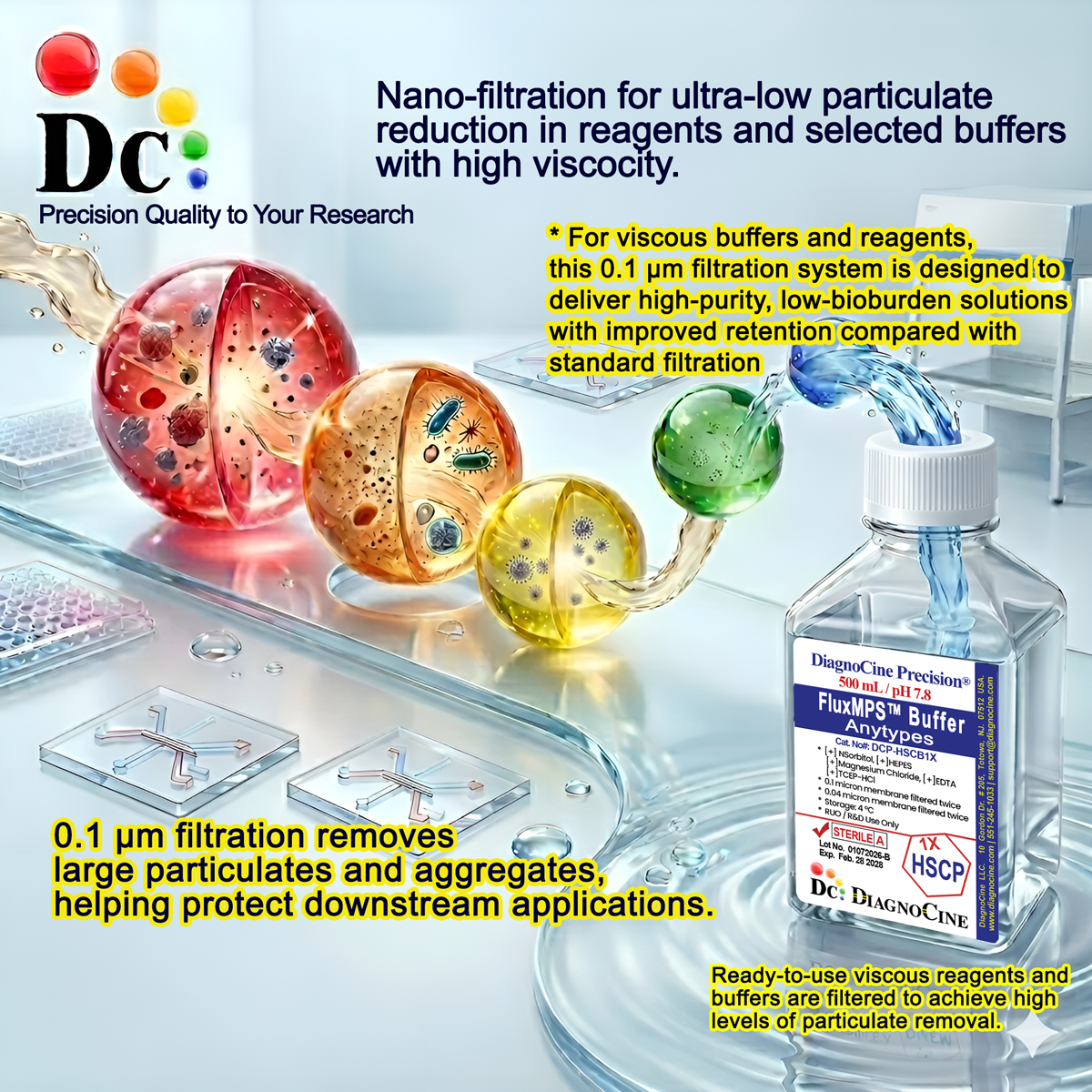
0.1 µm filtration removes large particulates and aggregates, helping protect downstream applications.
Precise, stable pH
Formulated at pH 7.5 with a defined phosphate-buffered saline composition for consistent, reproducible blocking performance lot to lot.
Ultrapure-grade water
Prepared with Ultrapure Type 1 water (18.2 MΩ·cm), meeting USP <85> water quality expectations for sensitive assay buffers.
Low background for imaging & assays
PVP-40 exhibits very low autofluorescence, reducing background noise in fluorescence detection channels compared with protein-based blockers.
Defined, traceable composition
Every salt and polymer component is formulated to a stated concentration, supporting reproducible blocking chemistry across experiments.
Customization on demand
Alternate concentrations, additional chemicals, compounds, proteins, supplements, or pH values can be formulated on request.
Single-stage 0.1 µm filtration
PBS Blocking Buffer with PVP is filtered through a 0.1 µm membrane for cell and molecular biology experiments.
- 1
0.1 µm Membrane Filtration
Removes large particulates and aggregates, helping protect downstream applications.
Filtration
0.1 µm filtration removes large particulates and aggregates, helping protect downstream applications.
© Diagnocine® — DCP-PVPPBS1X
Where PBS Blocking Buffer with PVP is used
A non-protein blocking buffer suited to immunoassays, sample handling, and imaging workflows where background reduction and reproducible chemistry matter.
Dot Blot & Protein Arrays
Blocks unoccupied binding sites on membranes and array surfaces to reduce non-specific antibody binding.
Wash, Dilution & Reconstitution
A defined-pH PBS base suitable for washing, diluting, and reconstituting samples ahead of downstream assays.
iPSC-Derived Model Handling
Non-protein, low-background buffer compatible with sensitive handling of iPSC-derived cell models.
Endothelial & Primary Cell Washing
Defined-composition PBS suitable for wash steps with endothelial and primary cell cultures.
ELISA, Blotting & Blocking
1% PVP-40 blocks non-specific binding on nitrocellulose and PVDF membranes and microplates, improving specificity in Western blotting and ELISA.
Microscopy & Optical Sensing
Very low PVP autofluorescence reduces background in fluorescence detection channels used in confocal and biosensor workflows.
Full specification summary
Measured and declared parameters for PBS Blocking Buffer with PVP.
| Parameter | Specification |
|---|---|
| Formulation / Composition | Phosphate-buffered saline with 1% PVP-40 |
| Appearance | Clear, colorless liquid |
| pH USP <791> | 7.5 |
| Sodium Chloride | 137 mM |
| Potassium Chloride | 2.7 mM |
| Sodium Phosphate Dibasic Anhydrous | 10.1 mM |
| Potassium Phosphate Monobasic Anhydrous | 1.76 mM |
| Parameter | Specification |
|---|---|
| Sterility | Nonsterile |
| DNase Activity | None detected (plasmid DNA, 18 hr incubation, room temperature) |
| RNase Activity | None detected (ribosomal RNA, 18 hr incubation, room temperature) |
| Water Quality | Ultrapure Type 1 water (18.2 MΩ·cm) |
| Manufacturing Standard ISO 13485 | ISO 13485-certified facilities |
| Parameter | Specification |
|---|---|
| Storage Temperature | 4°C |
| Shelf Life | 9 months |
| Parameter | Specification |
|---|---|
| Manufacturing QMS ISO 13485 | ISO 13485:2016 |
| Production Location | Diagnocine Precision, Totowa, New Jersey, USA |
| Intended Use | Research Use Only (RUO) |
Full composition
Every buffer salt and blocking polymer component, exactly as formulated in PBS Blocking Buffer with PVP.
| Component | CAS Number | Concentration |
|---|---|---|
| PVP-40 (Polyvinylpyrrolidone) | 9003-39-8 | 1% |
| Sodium Chloride (NaCl) | 7647-14-5 | 137 mM |
| Potassium Chloride (KCl) | 7447-40-7 | 2.7 mM |
| Sodium Phosphate Dibasic Anhydrous (Na2HPO4) | 7558-79-4 | 10.1 mM |
| Potassium Phosphate Monobasic Anhydrous (KH2PO4) | 7778-77-0 | 1.76 mM |
Manufacturing & compliance
PBS Blocking Buffer with PVP is manufactured, packaged, and tested under a controlled quality system.
ISO 13485:2016 QMS
Manufactured under ISO 13485-certified facilities that supply Diagnocine Precision.
Ultrapure Type 1 Water
Formulated with Ultrapure Type 1 water (18.2 MΩ·cm) as the aqueous base.
0.1 µm Filtration
0.1 µm filtration removes large particulates and aggregates, helping protect downstream applications.
Micro-Batch Precision
Final packaging, quality assurance, and testing are completed at the Diagnocine R&D and Quality Testing Center.
DNase / RNase Testing
Each lot is evaluated for DNase and RNase activity after 18 hr incubation with plasmid DNA and ribosomal RNA, respectively, at room temperature.
Sterility
Nonsterile.
Formulation Verification
Buffer salt and PVP-40 concentrations are formulated to the stated targets for each production lot.
Documentation / CoA
Certificates of Analysis document appearance, pH, and nuclease testing results for this product.
How DCP-PVPPBS1X compares
A snapshot of how this PVP-based blocking buffer compares with conventional protein-based and standard blocking buffer options.
| Parameter | DCP-PVPPBS1X | Conventional Protein-Based Blocker | Standard Blocking Buffer |
|---|---|---|---|
| Blocking chemistry | check_circle PVP-40 (non-protein) | cancel Protein-based (milk/BSA) | cancel Protein-based (milk/BSA) |
| Low autofluorescence | check_circle | cancel | cancel |
| DNase / RNase tested | check_circle | cancel | cancel |
| ISO 13485 manufacturing | check_circle | cancel | cancel |
| Immunoassay compatible | check_circle | check_circle | check_circle |
| Custom formulation available | check_circle | cancel | cancel |
Frequently asked questions
Common questions about PBS Blocking Buffer with PVP.
Supporting literature
Curated literature supporting the use of PVP-based blocking chemistry and phosphate-buffered saline in immunoassays.
- Bass, J.J. et al. An overview of technical considerations for Western blotting applications to physiological research. Scand J Med Sci Sports. 2017. doi:10.1111/sms.12702
- Mahmood, T. and Yang, P.C. Western blot: technique, theory, and trouble shooting. N Am J Med Sci. 2012. doi:10.4103/1947-2714.100998
- Aydin, S. A short history, principles, and types of ELISA, and our laboratory experience with peptide/protein analyses using ELISA. Peptides. 2015. doi:10.1016/j.peptides.2014.11.005
- Kurien, B.T. and Scofield, R.H. Western blotting. Methods. 2006. doi:10.1016/j.ymeth.2005.07.018
- Bewick, V., Cheek, L. and Ball, J. Statistics review: presenting and summarising data. Crit Care. 2003. doi:10.1186/cc2401
- Good, N.E. et al. Hydrogen ion buffers for biological research. Biochemistry. 1966. doi:10.1021/bi00866a011