


OmniFix Tissue Clearing Kit
FluxMPS™ OmniFix Tissue Clearing Kit
The FluxMPS™ OmniFix Tissue Clearing Kit (Cat. No. DCP-OTCK) is a ready-to-use, dual-solution optical clearing system for pre-fixed tissue samples. Both solutions are membrane-filtered through a 0.1 µm pore size, single pass, exceeding the particulate performance of standard 0.22 µm filtered reagents — supporting deep imaging, enhanced antibody penetration, and reproducible histological results across common fixation chemistries.
- 0.1 µm single-stage membrane filtration: Solution A and Solution B are each filtered through a 0.1-micron membrane once, exceeding the particulate performance of standard 0.22 µm filtered reagents.
- Broad fixation compatibility: Validated for EMA, PFA, formalin, ethanol/methanol, and glutaraldehyde-fixed tissue samples.
- Ready-to-use dual-solution system: Solution A (EZ View Solution) + Solution B (TF Lipid Removal Solution), pre-formulated to minimize preparation time and user error.
- Rapid optical transparency: Transforms opaque fixed tissue into optically clear samples while preserving structural integrity.
- Antigen & fluorophore preservation: Maintains protein epitopes and fluorescent protein signals for reliable immunostaining and advanced microscopy.
- Versatile imaging support: Compatible with confocal, light-sheet, and wide-field fluorescence microscopy, as well as standard histological analysis.
- ISO 13485:2016 manufacturing: Assembled, customized, and released at Diagnocine Precision, Totowa, New Jersey, USA.
- Customizable: pH, concentration, additives, and modified protocols available on request — contact support@diagnocine.com.
- Solution A (EZ View Solution)100 mL | pH 7.4 ± 0.04
- Solution B (TF Lipid Removal Solution)100 mL | pH 6–7
- AppearanceClear-colorless solution
- Filtration0.1 µm membrane, single pass
- Fill EnvironmentNon-sterile controlled environment
- StorageRoom Temperature
- Shelf Life — Solution A6 months
- Shelf Life — Solution B1 month
- Post-clearing (short-term)RT, up to 1 week in EZ View
- Post-clearing (long-term)RT, up to 70 days in EZ View
Engineered where conventional clearing methods fall short
Standard tissue clearing protocols require laborious solution preparation and often suffer from inconsistent clearing depth, signal loss, and limited fixation compatibility. The FluxMPS™ OmniFix Tissue Clearing Kit addresses these bottlenecks with a factory-formulated, 0.1 µm filtered dual-solution system that supports reproducible optical clarity across major fixation chemistries.
Low-Particulate Purity
A quadruple-stage train (0.1 µm ×2 + 0.04 µm ×2) reaches a 0.04 µm final cut-off, five times finer than the 0.22 µm membranes used for conventional sterile filtration.
Optical Transparency
The EZ View Solution (Solution A) transforms opaque fixed tissue into optically clear samples, supporting deep-volume confocal, light-sheet, and wide-field fluorescence imaging.
Broad Fixation Compatibility
Validated for EMA, PFA, formalin, ethanol/methanol, and glutaraldehyde-fixed tissues — one kit covers the majority of common histological fixation protocols without reformulation.[1]
Antigen & Fluorophore Preservation
Maintains protein epitopes and fluorescent protein signals throughout clearing, supporting downstream immunostaining, antibody labeling, and live-reporter readouts.[2]
Ready-to-Use Dual-Solution System
Pre-formulated Solution A and Solution B reduce the preparation errors common with manually mixed clearing reagents, supporting reproducibility across experiments.
Customization on Demand
Alternative concentrations, pH values, chemical additions, or modified protocols are available. Contact support@diagnocine.com with your specifications.
Filtration
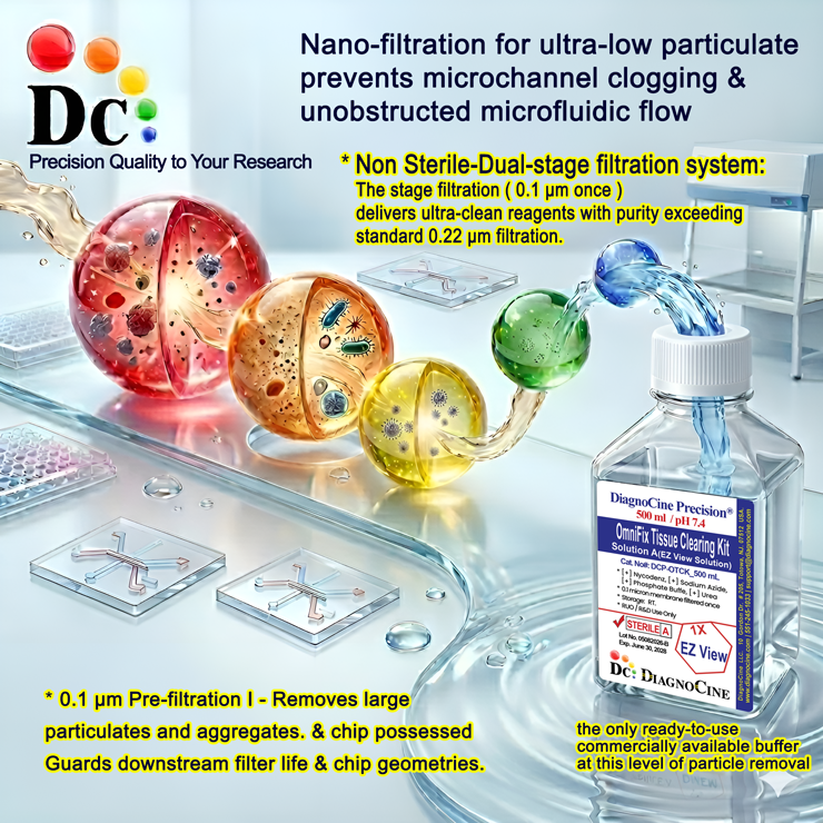
Both solutions in the OmniFix Tissue Clearing Kit are filtered through a 0.1-micron membrane, once. This single-pass 0.1 µm filtration exceeds the particulate-reduction performance of standard 0.22 µm filtered reagents, reducing the sub-micron particulate burden that can compromise optical clarity during imaging. The process is performed in a controlled, non-sterile manufacturing environment under an ISO 13485:2016 quality management system.
-
1
0.1 µmMembrane Filtration
Both Solution A (EZ View Solution) and Solution B (TF Lipid Removal Solution) are passed through a 0.1 µm membrane filter exactly once. This removes large particulates, aggregates, and sub-micron debris that could otherwise affect refractive index uniformity and optical clarity during imaging. The 0.1 µm pore size retains material that a conventional 0.22 µm filter passes, protecting sensitive detector optics and microfluidic chip geometries.
Filtration architecture vs. standard 0.22 µm filtered reagents
A single pass through a 0.1 µm membrane captures particle populations that pass freely through a 0.22 µm filter. In optical clearing workflows, sub-micron particulates can create scattering artifacts that reduce effective imaging depth and resolution. OmniFix reagents apply 0.1 µm filtration to reduce this particulate load relative to conventionally filtered reagents.
pore size
stage
© Diagnocine® — DCP-OTCK
Optical clearing for next-generation tissue imaging
OmniFix is designed for researchers requiring reliable, deep-volume imaging of fixed tissue samples across diverse biological and pathological workflows. The kit supports immunofluorescence, 3D reconstruction, and multi-modal microscopy on a range of tissue types and fixation protocols.
Microfluidic & Chip-Compatible Use
The reduced particulate profile of OmniFix reagents (0.1 µm filtered) supports use with microfluidic perfusion systems and organ-on-a-chip (OoC) platforms, where particulate accumulation can occlude microchannels or degrade sensor signals. A finer, 0.01 µm (10 nm) ultra-filtered variant is available on inquiry for automated bioreactors and robotic perfusion systems.
- Reduced Particulate Load: 0.1 µm filtration removes debris above threshold, reducing the risk of microchannel occlusion in chip-integrated clearing workflows.
- Valve & Sensor Protection: Lower particle counts help protect microfluidic valves, pressure sensors, and optical detection elements in automated systems.
- Reagent Stability: Stable reagent chemistry helps prevent precipitation or viscosity drift that could interrupt continuous-flow clearing protocols.
Inquiry Required: The 0.01 µm (10 nm) ultra-filtered OmniFix variant is available on special request. Contact support@diagnocine.com with your system specifications.
3D Volumetric Tissue Imaging
OmniFix provides the refractive index matching and optical transparency required for undistorted light-sheet illumination throughout thick tissue volumes, supporting whole-organ 3D reconstruction.[3]
Deep-Volume Confocal Imaging
Reduces light-scattering artifacts to support confocal z-stack acquisition at depths less accessible with non-cleared tissues. Compatible with multi-photon excitation and super-resolution modalities.[4]
Antibody Labeling & IF Staining
Preserves antigen accessibility and fluorescent protein integrity throughout the clearing pipeline, supporting immunofluorescence, secondary antibody penetration, and multiplex staining in thick sections.[2]
3D Anatomical Reconstruction
Supports 3D rendering of complex tissue architectures including neural circuits, vascular networks, and organoid structures from fixed specimens without physical sectioning artifacts.
Morphology & Pathology Studies
Cleared specimens retain structural features suitable for morphological assessment, disease staging, and cellular architecture analysis in research pathology workflows.[5]
OoC / MPS Tissue Imaging
Reduced-particulate reagents (0.1 µm filtered) are suitable for on-chip tissue clearing in organ-on-a-chip (OoC), microphysiological system (MPS), and tissue-on-a-chip (ToC) platforms where low-particulate reagents are required.
Analytical & quality specifications
All parameters are measured per lot and documented in the Certificate of Analysis. Contact support@diagnocine.com to request a CoA for a specific lot number.
| Parameter | Specification |
|---|---|
| Formulation | Nycodenz, Urea, Sodium Azide, Phosphate Buffer |
| Appearance | Clear-colorless solution |
| pH USP <791> | 7.4 ± 0.04 at 1X |
| RI application | Refractive index matching for optical clearing |
| Protocol role | Step C — RI Matching (24 h incubation) |
| Imaging note | Maintain tissue in EZ View during microscopy |
| Parameter | Specification |
|---|---|
| Formulation | Tetrahydrofuran (THF), Ultrapure Water |
| Appearance | Clear-colorless solution |
| pH USP <791> | 6–7 at 1X |
| THF content | Approximately 50% THF in sterile water (working concentration) |
| Protocol role | Step A — Lipid Removal (16 h incubation) |
| Safety | Use in chemical fume hood; chemical waste disposal |
| Parameter | Specification |
|---|---|
| Filtration pore size | 0.1 µm membrane |
| Filtration stages | 1 (single pass) |
| Fill environment | Non-sterile controlled manufacturing environment |
| Sterility declaration | Filtered through a 0.1-micron membrane once |
| Manufacturing ISO 13485 | ISO 13485:2016 quality management system |
| Water quality (Sol. B) | Ultrapure Water |
| Parameter | Specification |
|---|---|
| Storage temperature | Room Temperature |
| Shelf life — Solution A | 6 months |
| Shelf life — Solution B | 1 month |
| Expiry date | Use before expiry date on product label |
| Post-clearing short-term | Store in EZ View at RT, up to 1 week |
| Post-clearing long-term | Store in EZ View at RT, up to 70 days |
Full composition
The OmniFix Tissue Clearing Kit contains two pre-formulated solutions. Ingredient identities and working concentrations are proprietary to Diagnocine. CAS numbers are provided for informational reference. Nucleic acids from OmniFix-cleared tissues will be fragmented rather than intact — the degree of fragmentation depends on fixation duration and must be optimized per experiment.
| Ingredient | CAS Number | Concentration |
|---|---|---|
| SOLUTION A — EZ VIEW SOLUTION (pH 7.4 ± 0.04) | ||
| Nycodenz | 66108-95-0 | Proprietary |
| Urea | 57-13-6 | Proprietary |
| Sodium Azide | 26628-22-8 | Proprietary |
| Phosphate Buffer | N/A (mixture) | Proprietary |
| Ingredient | CAS Number | Concentration |
|---|---|---|
| SOLUTION B — TF LIPID REMOVAL SOLUTION (pH 6–7) | ||
| Tetrahydrofuran (THF) | 109-99-9 | Proprietary (approx. 50% working) |
| Ultrapure Water | 7732-18-5 | Proprietary |
| Fixation Type | EZ Clear Compatibility | Notes / Considerations |
|---|---|---|
| FIXATION COMPATIBILITY TABLE | ||
| EMA (ethanol-methanol-acetic acid) | High | Original validated use |
| PFA (Paraformaldehyde) | Moderate–High | May need longer clearing; FP preserved |
| Formalin | Moderate | Slower clearing; some opacity may remain |
| Ethanol / Methanol | High | Very compatible, similar to EMA |
| Glutaraldehyde | Low | Strong crosslinking limits clearing |
EMA-fixed tissue clearing — complete EZ Clear protocol
Full three-step tissue processing sequence for EMA-fixed samples. Adapt incubation times as needed for other fixation types per the compatibility table above.
Step A: Lipid Removal (16 hours)
Using Solution B (TF Lipid Removal Solution) in a chemical fume hood:
- Transfer fixed tissue to a glass scintillation vial (20 mL capacity).
- Add 20 mL THF solution (approximately 50% THF in sterile water).
- Cover with foil to protect from light.
- Place on an orbital shaker in the chemical fume hood.
- Incubate 16 hours at room temperature with gentle rocking.
Step B: Washing (4 hours)
- Remove THF solution — dispose in a chemical waste container.
- Rinse with sterile Milli-Q water 4 times, 1 hour each.
- Use 20 mL water per wash, with gentle rocking.
- Final ventilation — open vial in the hood for 10 minutes to remove residual THF.
Step C: Refractive Index Matching (24 hours)
Using Solution A (EZ View Solution):
- Transfer tissue to a clean vial or imaging chamber.
- Remove all PBS/water completely.
- Add 5 mL EZ View solution (or sufficient to cover the tissue).
- Protect from light with foil wrapping.
- Incubate 24 hours at room temperature with gentle rocking.
- Sample is ready for imaging — maintain in EZ View during microscopy.
Troubleshooting Guidelines
Manufacturing standards & compliance
The OmniFix Tissue Clearing Kit is manufactured under ISO 13485-certified quality system standards. Final packaging, quality assurance, and testing are performed at the Diagnocine R&D and Quality Testing Center. All customization and assembly are completed at Diagnocine Precision in Totowa, New Jersey, USA.
ISO 13485:2016 QMS
Manufactured under a full ISO 13485:2016 quality management system. Products are released only after QC testing at the Diagnocine R&D and Quality Testing Center.
0.1 µm Membrane Filtration
Both solutions are filtered through a 0.1-micron membrane (single pass) in a controlled manufacturing environment, reducing particulate load relative to 0.22 µm filtered reagents.
Diagnocine Precision Assembly
All final kit assembly, labeling, customization, and quality release are performed at Diagnocine Precision, Totowa, New Jersey, USA under controlled laboratory conditions.
Lot-Specific CoA
Each lot is accompanied by a Certificate of Analysis documenting pH, appearance, filtration grade, and shelf-life validation. Contact support@diagnocine.com for a lot-specific CoA.
pH Verification
Solution A: pH 7.4 ± 0.04 at 1X (per lot CoA). Solution B: pH 6–7. Measured by calibrated pH meter per established SOP.
Appearance QC
Both solutions must present as a clear-colorless liquid on inspection. Turbidity, color, or particulate contamination constitutes a lot-failure event.
Filtration Verification
0.1 µm membrane filtration is applied to every production batch. Filter integrity is tested before and after filtration per an internal protocol aligned with ISO 13485 requirements.
Documentation & Traceability
Full batch records including raw material traceability, filtration logs, QC results, and CoA are maintained. Regulatory documentation is available upon request.
How DCP-OTCK compares to conventional clearing methods
OmniFix benchmarked against commonly used manual and commercial tissue clearing approaches, highlighting differences in purity, compatibility, and workflow convenience.
| Parameter | DCP-OTCK (FluxMPS™ OmniFix) | Manual iDISCO / BABB Method | Generic Commercial Clearing Kit (0.22 µm) |
|---|---|---|---|
| Reagent format | Ready-to-use pre-formulated dual solution | Manual preparation required | Semi-prepared; some mixing required |
| Filtration grade | Single-stage 0.1 µm filtration (not the Microfluidics Suitable 0.04 µm tier) | Not specified | Single-pass 0.22 µm |
| Filtration pore size | 0.1 µm | Typically unfiltered or 0.22 µm | 0.22 µm standard |
| Filtration stages | 1 (single pass) | Not specified | 1 (single pass) |
| EMA-fixed tissue compatibility | check_circle High | cancel Not validated | cancel Not validated |
| PFA compatibility | check_circle Moderate–High | check_circle Yes (primary use) | check_circle Yes |
| Formalin compatibility | check_circle Moderate | cancel Limited | cancel Limited |
| Antigen/fluorophore preservation | check_circle Yes | check_circle Variable | check_circle Variable |
| Manufacturing QMS | ISO 13485:2016 | No certification | Varies by supplier |
| Custom formulation | check_circle Available on request | cancel No | cancel No |
Frequently asked questions
Common questions about the OmniFix Tissue Clearing Kit (DCP-OTCK).
Supporting literature
Curated peer-reviewed references on tissue optical clearing, lipid-extraction protocols, refractive index matching, and advanced fluorescence microscopy of cleared specimens.
- Erturk A, et al. Three-dimensional imaging of solvent-cleared organs using 3DISCO. Nature Protocols. 2012;7(11):1983–1995.doi:10.1038/nprot.2012.119
- Renier N, et al. iDISCO: a simple, rapid method to immunolabel large tissue samples for volume imaging. Cell. 2014;159(4):896–910.doi:10.1016/j.cell.2014.10.010
- Tomer R, et al. Advanced CLARITY for rapid and high-resolution imaging of intact tissues. Nature Protocols. 2014;9(7):1682–1697.doi:10.1038/nprot.2014.123
- Susaki EA, et al. Whole-brain imaging with single-cell resolution using chemical cocktails and computational analysis. Cell. 2014;157(3):726–739.doi:10.1016/j.cell.2014.03.042
- Hama H, et al. Scale: a chemical approach for fluorescence imaging and reconstruction of transparent mouse brain. Nature Neuroscience. 2011;14(11):1481–1488.doi:10.1038/nn.2928
- Pan C, et al. Shrinkage-mediated imaging of entire organs and organisms using uDISCO. Nature Methods. 2016;13(10):859–867.doi:10.1038/nmeth.3964
- Zhao S, et al. Cellular and molecular probing of intact human organs. Cell. 2020;180(4):796–812.doi:10.1016/j.cell.2020.01.030
- Ariel P. A beginner's guide to tissue clearing. International Journal of Biochemistry & Cell Biology. 2017;84:35–39.doi:10.1016/j.biocel.2016.12.009
- Marx V. Optimizing probes to image cleared tissue. Nature Methods. 2016;13(3):205–208.doi:10.1038/nmeth.3774
- Vigouroux RJ, Belle M, Chedotal A. Neuroscience in the third dimension: shedding new light on the brain with tissue clearing. Molecular Brain. 2017;10(1):33.doi:10.1186/s13041-017-0314-y