HistoBright, Tissue Structure Fluorescent Dye
Product#: FNK-FDV-0051
$538.20
Availability:
Ships in 1-2 Weeks
HistoBright (Tissue Structure Fluorescent Dye)
Cat. No.: FNK-FDV-0051
Description
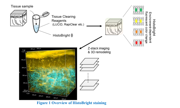
HistoBright is an innovative tissue structure fluorescent dye designed for high-contrast visualization and 3D imaging of biological tissues. This environment-responsive solvatochromic dye changes its fluorescence properties based on the polarity of the surrounding microenvironment, providing a versatile tool for tissue imaging. HistoBright is particularly effective for deep-tissue imaging when combined with tissue-clearing techniques, enabling researchers to analyze thick tissue blocks without disrupting their pathophysiological structures.
Key Features:
1. Solvatochromic properties: Fluorescent emission shifts from green to far-red depending on the solvent polarity, enhancing contrast and revealing microenvironmental details.
2. Wide excitation/emission range: Excitation at 350–500 nm (maximum ~410 nm) and emission between 500–800 nm, making it compatible with 405–488 nm lasers for confocal microscopy and 960–1100 nm lasers for two-photon microscopy.
3. High tissue permeability: Efficiently penetrates tissues for uniform staining.
4. Dual compatibility: Suitable for both one-photon and two-photon microscopy.
5. Post-staining flexibility: Allows subsequent use of conventional histological techniques like Hematoxylin and Eosin (HE) staining after fluorescent observation.
6. Durable formulation: Molecular weight of 381.5 g/mol; supplied as a yellow liquid soluble in DMSO.
Applications:
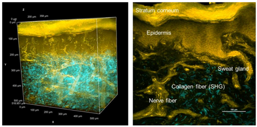
1. 3D tissue imaging: Enables visualization of thick tissue blocks while preserving structural integrity, especially when paired with clearing methods like LUCID or RapiClear.
2. Confocal and two-photon microscopy: Ideal for high-resolution imaging of biological membranes and tissue structures.
3. Deep-tissue analysis: Facilitates spatial structural analysis of intact tissues without requiring thin slicing.
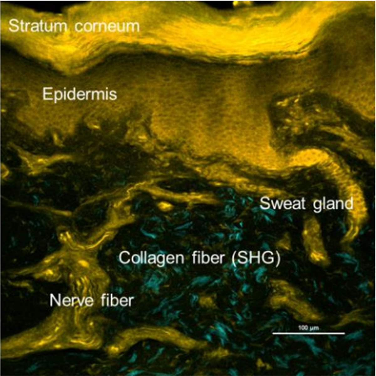
4. SHG imaging compatibility: Simultaneously visualizes collagen fibers via second harmonic generation without additional staining.
Advantages:
1. Enhanced visualization: Solvatochromic fluorescence provides high contrast by adapting to local environmental changes, making subtle tissue structures more discernible.
2. Versatile spectral range: Wide excitation/emission compatibility allows integration into various imaging systems.
3. 3D structural preservation: Eliminates the need for thin slicing, maintaining native tissue architecture during analysis.
4. Multiplexing potential: Compatible with multiple laser systems and other fluorescence-based probes for multi-color imaging.
5. Ease of use: Can be seamlessly incorporated into existing workflows for confocal or two-photon microscopy.
Reconstitution and Storage
1. Reconstitution: Prepare stock solution in 100% DMSO.
2. Storage conditions:
-Store powder at -20°C.
-After reconstitution, aliquot and store at -20°C to prevent repeated freeze-thaw cycles.
-Protect from light to maintain stability.
HistoBright represents a groundbreaking advancement in tissue imaging, offering researchers the ability to visualize complex 3D structures with exceptional clarity. Its environment-responsive fluorescence, combined with compatibility across various microscopy platforms, makes it an indispensable tool for applications in cell biology, histology, and biomedical research.
Specification
Formulation: C27H27NO
Molecular weight: 381.5 g/mol
Solubility: Soluble in DMSO
Fluorescent characteristics:
- Ex. 350-500 nm (maximum ~410 nm) Compatible with 405-488 nm laser (confocal microscopy), 960-1100 nm laser (two-photon microscopy)
- Em. 500-800 nm (depending on solvents)
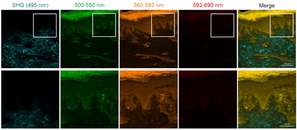
FNA HistoBright FNA HistoBright epidermis imaging
FNA HistoBright FNA HistoBright dermis imaging
Description
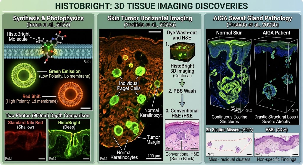
Tissue structures are historically visualized by staining with various colorimetric dyes such as Hematoxylin and Eosin (HE) stains. These conventional colorimetric stains generally require thinly sliced tissue sections and are unsuitable for thick tissue blocks. However, the preparation of thinly sliced tissues loses the 3D structural information. To reconstitute 3D information, a lot of additional thin slices and stainings are required. HistoBright (original compound name; PC in Ref.1) is originally synthesized by Dr. Yosuke Niko, Kochi University, and Dr. Takeshi Imamura, Dr. Masamoto Murakami, and Dr. Ryosuke Kawakami, Ehime University, promotes its biological 3D imaging application. HistoBright is a novel solvatochromic fluorescent dye that changes fluorescent colors in response to solvents or microenvironments and has a high affinity to various biological membranes. The combination of two features of HistoBright contributes to clear and bright imaging of tissue structures. As HistoBright can be excited by not only one-photon but also two-photon, HistoBright is compatible with two-photon microscopy and useful for tissue imaging. Furthermore, HistoBright, coupled with several tissue-clearing methods such as LUCID and RapiClear, which do not require lipid removal, contributes to analyzing 3D deep tissue imaging of thick tissue slices or blocks, etc., without breaking pathophysiological tissue structures. HistoBright is an innovative fluorescent dye for the visualization of 3D tissue structures.
Reconstitution and Storage
- Reconstitution: Stock solution recommended concentration 1 mM in 100% DMSO.
- Storage (powder): Store powder at -20°C.
- Storage (solution): After reconstitution in DMSO, aliquot and store at -20°C. Avoid repeated freeze-thaw cycles.
How to use
General procedure for tissue staining
*This procedure is an example of HistoBright
- Tissue samples were excised and then fixed in freshly prepared paraformaldehyde.
- Tissue blocks or thick slices prepared by any appropriate methods.
- Stock solution of HistoBright (1 mM) was mixed in tissue clearing solution (LUCID or RapiClear etc.) to prepare the staining and clearing solution (hereafter called Master mix). The initial recommendation of HistoBright concentration is 10 uM.
- Immerse tissue blocks/sections in the Master mix for over 48 hours at room temperature.
- After staining tissue blocks/sections, image by confocal laser microscopy or two-photon microscopy.
- After fluorescence imaging, immerse the tissue block/section in phosphate-buffered saline (PBS) to remove residual HistoBright™ dye.
- Proceed to traditional H&E staining or other histological protocols after PBS washing.
Reference Data
| Solvatochromic property of HistoBright | |
| Absorbance and fluorescent spectrum of HistoBright in various solvents. While the absorbance spectrum of HistoBright is not sensitive to solvent polarity, HistoBright changes fluorescent color from green to far-red depending on solvent polarity. Under a low polarity environment, HistoBright emits green color. On the other hand, under a high polarity environment, HistoBright emits red to far-red with low quantum yield. Quantum yields are 0.82 (Toluene), 0.77 (EtOAc), 0.83 (CHCl3), 0.65 (MeCN), 0.20 (EtOH), and 0.07 (MeOH). | 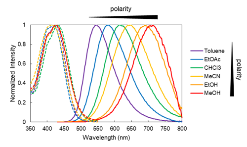 |
| Fluorescent spectrum on a liposome membrane model | |
| HistoBright rapidly accumulates in lipid membranes and emits different fluorescent colors in response to the membrane environments. On the liquid order (Lo) model liposome (sphingomyelin (SM)/cholesterol (Chol)), HistoBright emits a maximum of around 575 nm fluorescence with a high quantum yield value (0.72). On the other hand, HistoBright on the liquid disorder (Ld) model liposome (DOPC) exhibits significantly broad and relatively weak fluorescence (quantum yield 0.37), and the maximum wavelength is red-shifted by approximately 30 nm compared to the Lo model. | 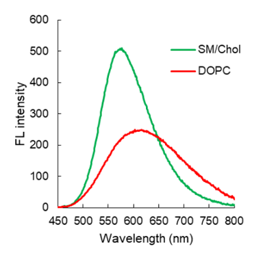 |
Application data
| Two-photon fluorescence microscopy imaging of human normal skin tissue | |
| After fixation of human skin tissue blocks with 4% paraformaldehyde/PBS, 500 um thick slides were prepared and treated with the LUCID clearing reagent and 10 uM HistoBright for 76 hours. The tissue slides were observed by two-photon fluorescence microscopy (Ex. 960 nm, Em. cyan channel: 492 nm as SHG, green channel: 500-550 nm, orange channel: 560-593 nm, and red channel: 593-690 nm). The merged image was obtained from four channels. | |
 |
|
| 3D images were reconstructed from Z-stack sequences (step size, 5 um) of 2D images | |
 |
|
| Comparison between confocal microscopy and two-photon microscopy imaging | |
| After fixation of human skin tissue blocks with 4% paraformaldehyde/PBS, 500 um thick slides were prepared and treated with the RapiClear clearing reagent and 10 uM HistoBright for 72 hours. Tissues were observed by confocal microscopy (488 nm laser) and two-photon microscopy (960 nm laser). | 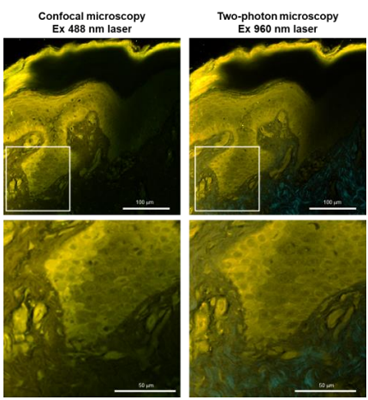 |
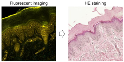
| HE staining after HistoBright fluorescent imaging | |
| After fluorescent imaging, the stained tissue block was washed with PBS. Thin tissue slices were prepared from the tissue block and re-stained with the conventional HE staining procedure. | |
 |
|
Notes
- All spectrum data were obtained by Dr. Yosuke Niko, Kochi University.
- All cell imaging data were obtained by Dr. Ryosuke Kawakami, Ehime University.
Reference
- Inoue et al.,J. Mater. Chem. B, 10, 1641-1649 (2022) Synthesis and photophysical properties of a new push-pull pyrene dye with green-to-far-red emission and its application to human cellular and skin tissue imaging
- Yoshida, S., Kawakami, R., Niko, Y., Yatsuzuka, K., Tsuda, T., Mori, H., Muto, J., Shiraishi, K., Imamura, T., Fujisawa, Y., & Murakami, M. (2025). Horizontal imaging of skin tumors using advanced fluorescence techniques for detecting individual malignant cells. Acta Histochemica et Cytochemica, 58(2), 81–92. https://doi.org/10.1267/ahc.24-00059
- Yoshida S, Kawakami R, Niko Y, Fujisawa Y, Murakami M. New Characteristics of Eccrine Sweat Glands in Acquired Idiopathic Generalised Anhidrosis as Determined via Three-Dimensional Fluorescence Imaging of Cleared Skin Tissue. Exp Dermatol. 2025 Mar;34(3):e70038.
Summary Comparison Table
| Publication | Primary Focus | Key Microscopy Technique | Major Achievement with HistoBright |
| Inoue et al. (2022) | Chemical synthesis and photophysical characterization. | Two-Photon Microscopy (960 nm excitation) | Proved high polarity-responsiveness, high quantum yield, and superior depth penetration over Nile Red. |
| Yoshida et al. (2025a) | Horizontal skin tumor edge mapping and single-cell tracking. | Confocal (488 nm) & Two-Photon | Pinpointed single malignant cells in thick tissue; demonstrated complete dye wash-out for subsequent H&E staining. |
| Yoshida et al. (2025b) | Pathology of Acquired Idiopathic Generalized Anhidrosis (AIGA). | Three-Dimensional Two-Photon |