HemeGREEN, Intracellular Labile Heme Detection Reagent
HemeGREEN, Intracellular Labile Heme Detection Reagent
Cat. No.: FNK-FDV-0056
Size: 0.1mg
Storage: -20°C
HemeGREEN™ is a fluorescent probe that enables visualization of intracellular labile heme in living cells. It exhibits high selectivity for labile heme over other reactive species such as ferrous iron (Fe2+) and heme tightly bound to proteins.
Intracellular Labile Heme Analysis
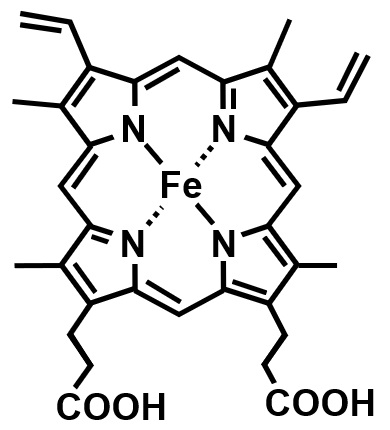 |
Heme is a coordination compound of protoporphyrin IX and an iron ion at its center. In bioligical systems, it plays a crucial role as a cofactor of heme proteins, such as oxygen-transport/storage proteins like hemoglobin and myoglobin, and metabolic or redox proteins like peroxidases and cytochrome P450. While most cellular heme exists tightly bound within heme proteins, a small amount of labile (unbound) heme also exists and has recently attracted attention. Several heme-responsive transcription factors, such as Bach2, have been identified, implicating labile heme in cellular differentiation, circadian rhythm, and disease pathogenesis. Due to its high lipophilicity and redox reactivity compared to free iron ions, labile heme is also thought to contribute to oxidative damage of biomolecules. However, analytical tools for detecting and visualizing intracellular labile heme have been limited, and its dynamics remain poorly understood. HemeGREEN™ is a small molecule fluorescent probe to enable selective detection of intracellular labile heme. |
Principle
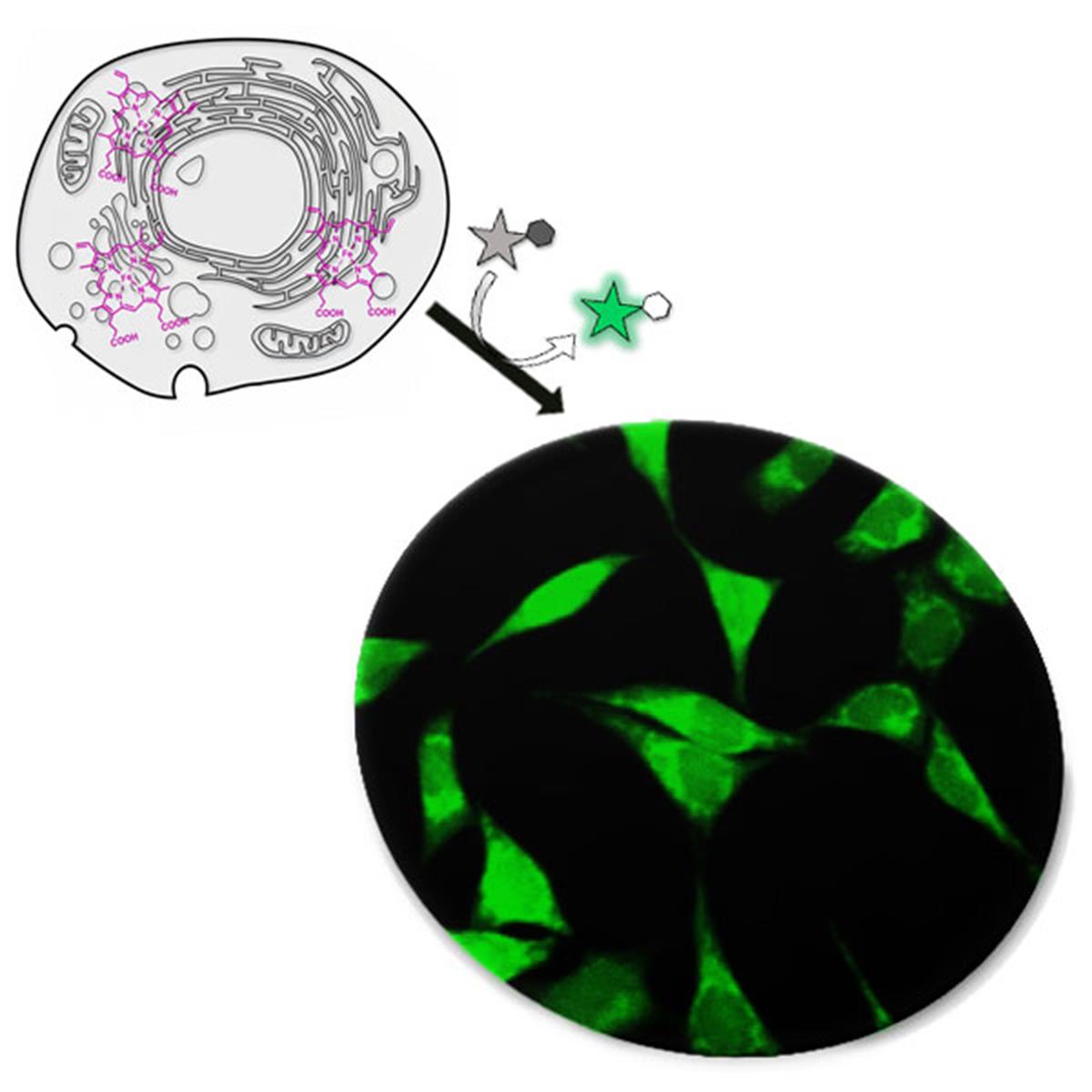
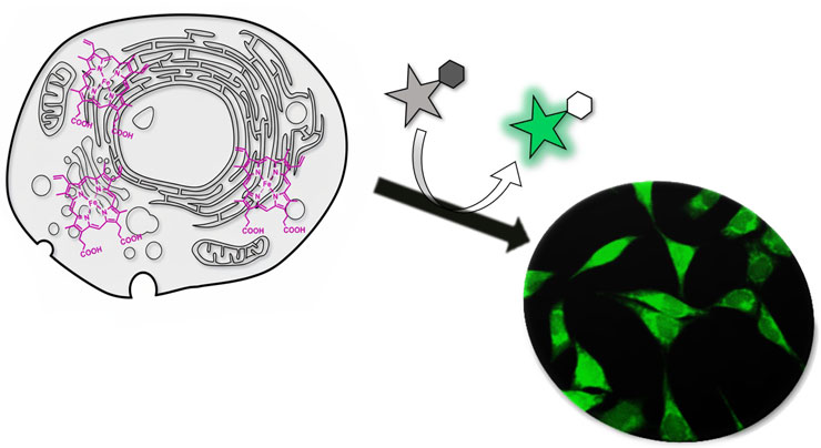
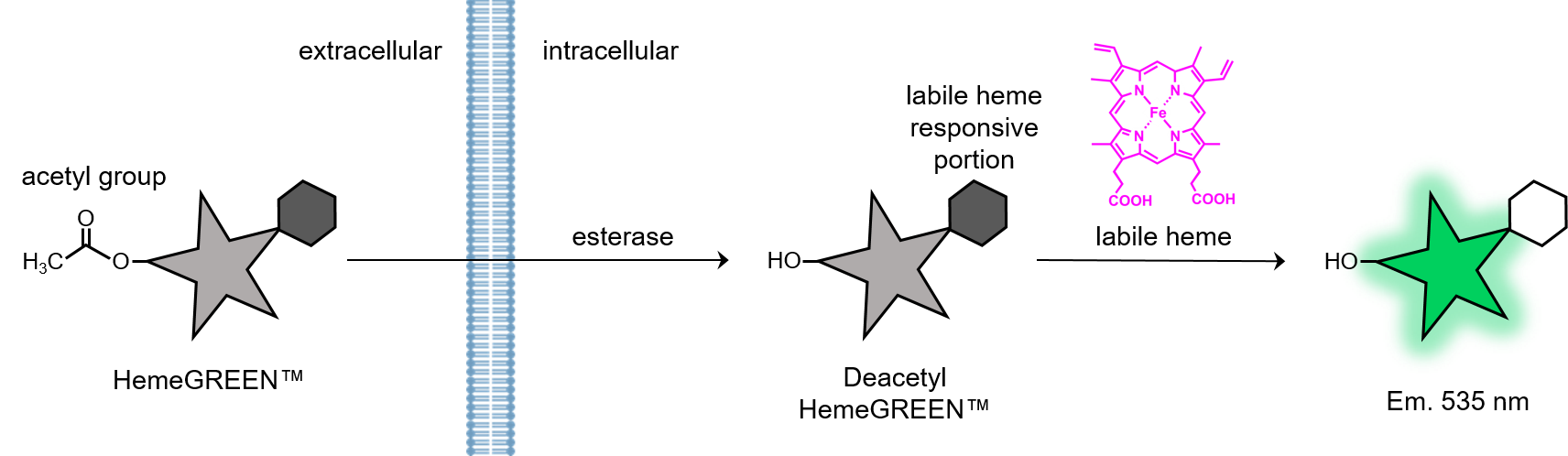
HemeGREEN™ consists of a Rhodol fluorophore with acetyl groups for enhancing cell permeability and a heme-responsive moiety. It exhibits almost no fluorescence in its native form. Once inside the cell, the acetyl groups are cleaved by intracellular esterases to yield deacetylated HemeGREEN™, which still does not fluoresce unless labile heme is present. In the presence of labile heme, the probe reacts with the heme to emit green fluorescence with a maximum emission at 535 nm.
※ HemeGREEN™ is designed exclusively for live-cell imaging and is not suitable for in vitro assays.
Features
- Fluoresces upon reaction with intracellular labile heme, allowing direct visualization.
- Highly selective for labile heme; minimal response to other metal ions or heme bound to proteins.
- Cell-permeable; spontaneously taken up by cells by simple addition to culture medium.
- Low cytotoxicity at the recommended working concentration (10 μM).
- Suitable for imaging and analysing labile heme dynamics in living cells.
- Molecular formula: C27H21F2NO6
- Molecular weight: 493.46
- Solubility: Soluble in DMSO
- Ex/Em: 490/535 nm (compatible with standard FITC filter sets)
Reference Data
Fluorescence Spectrum
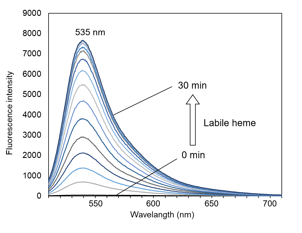 |
※ Data shown are for deacetylated HemeGREEN™; not for the intact acetylated form. Deacetylated HemeGREEN™ (0.2 μM) was dissolved in HEPES buffer (pH 7.4) containing 100 μM GSH, and 1 μM Hemin was added. Fluorescence changes were monitored over time. ※ Excitation wavelength: 490 nm ※ GSH reduces Fe(III) in Hemin to Fe(II), generating labile heme that reacts with HemeGREEN™. |
Selectivity for Labile Heme
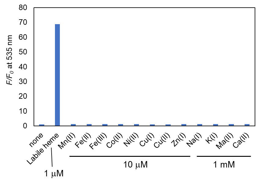 |
Deacetylated HemeGREEN™ (0.2 μM) was incubated with various metal ions in HEPES buffer (pH 7.4) at room temperature for 30 minutes in a 96-well plate. Fluorescence was measured using a plate reader with excitation at 480 nm and emission at 530 nm. ※ “Labile heme” condition corresponds to Hemin reduced with GSH. |
Assay Overview
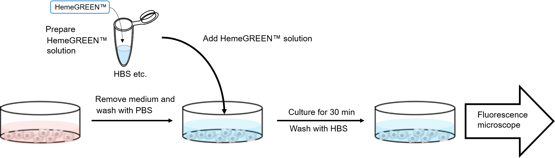
1. Prepare HemeGREEN™ working solution (final 10 μM) in HBS or equivalent buffer.
2. Remove culture medium and wash cells with PBS. 3. Add HemeGREEN™ solution to cells.
4. Incubate for 30 minutes.
5. Wash once with HBS or equivalent buffer.
※ Optimize probe concentration and incubation time depending on the cell type.
※ Fluorescence dye may diffuse over time; imaging is recommended immediately after washing.
Example Data
Monitoring Labile Heme Under Modulation of Heme Biosynthesis and Degradation
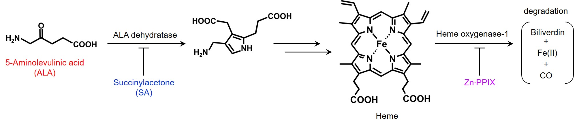
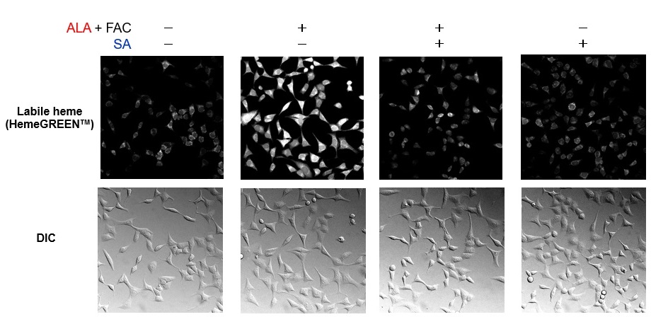
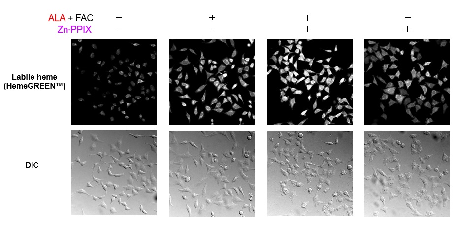
HeLa cells were treated overnight with 1 mM 5-aminolevulinic acid (ALA) and 10 μM ferric ammonium citrate (FAC) to promote heme biosynthesis. In parallel, cells were co-treated with 1 mM succinylacetone (SA), an inhibitor of ALA dehydratase, and 1 μM ZnPPIX, an inhibitor of heme oxygenase-1. The following day, cells were stained with HemeGREEN™ for 30 minutes and observed by confocal microscopy. ALA and FAC increased fluorescence, indicating enhanced labile heme levels. This signal was suppressed by SA, suggesting inhibition of heme biosynthesis. Co-treatment with ZnPPIX further enhanced fluorescence, indicating inhibition of heme degradation and accumulation of labile heme.
 |
 |
 |
Evaluation of NO-Induced Labile Heme Increase
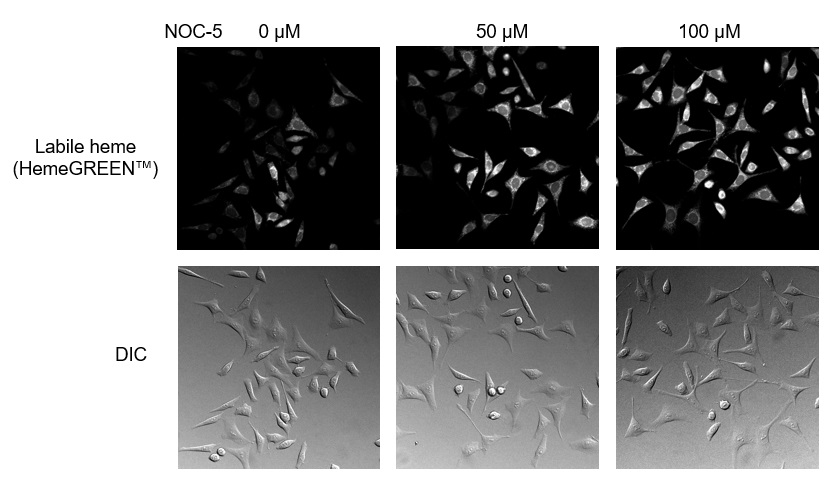
It has been known that NO-treatment of heme-binding proteins causes transient release of heme from the proteins. HemeGREEN™ detected this transient change of labile heme in living cells. HeLa cells were incubated with the NO donor NOC-5 for 20 minutes, followed by staining with HemeGREEN™ for 30 minutes. Confocal microscopy revealed a concentration-dependent increase in fluorescence, indicating that NO increased intracellular labile heme levels.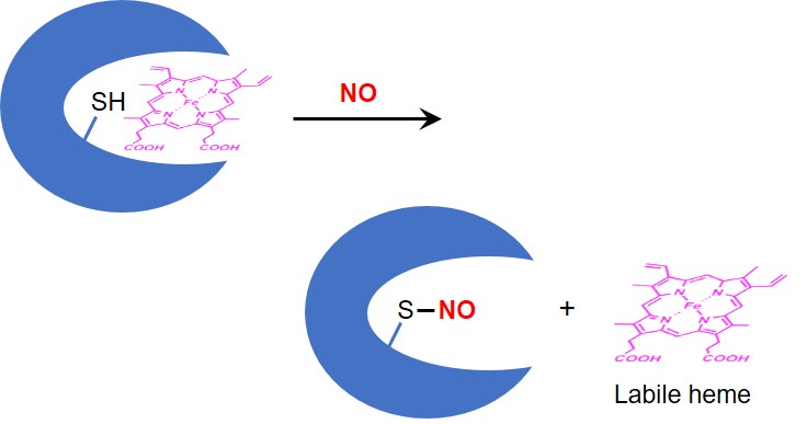 |
 |
Monitoring Labile Heme Levels During Ferroptosis
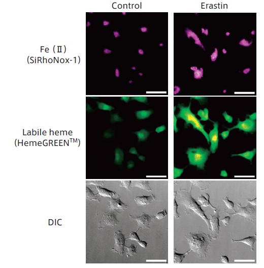 |
HT1080 cells were treated with the ferroptosis inducer erastin for 6 hours, then co-stained with HemeGREEN™ and the Fe(II) probe SiRhoNox-1. Confocal imaging revealed that both Fe(II) and labile heme levels increased simultaneously during ferroptosis induction. |
References
Kawai, K., et al., “Molecular Imaging of Labile Heme in Living Cells Using a Small Molecule Fluorescent Probe”, J. Am. Chem. Soc., 144 (9), 3793–3803 (2022). [PMID:35133144]