PBS Blocking Buffer with Gelatin (1%)
PBS Blocking Buffer with Gelatin (1%)
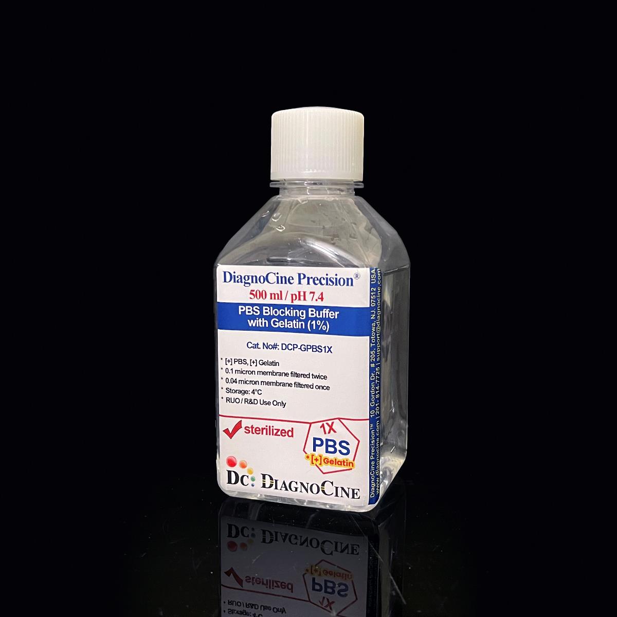
PBS Blocking Buffer with Gelatin (1%) is a research-grade, 0.1 µm-filtered PBS-based blocking reagent formulated for Western blotting, ELISA, and other antibody-based immunoassays. Built on a stable pH 7.4 phosphate-buffered saline base with 1% gelatin, it forms an effective blocking matrix over non-specific binding sites on nitrocellulose, nylon, and charge-modified nylon membranes, supporting low-background detection across cell and molecular biology workflows.
- 0.1 µm filtration removes large particulates and aggregates, helping protect downstream applications.
- Nonsterile; confirmed free of detectable DNase and RNase activity after 18-hour challenge incubation
- Stable pH 7.4 phosphate-buffered saline (PBS) base
- 1% gelatin blocking matrix, effective for protein-protein interaction blocking
- Compatible with nitrocellulose, nylon, and charge-modified nylon membranes (not recommended for PVDF membranes)
- Does not cross-react with mammalian antibodies
- Manufactured under ISO 13485:2016-certified facilities
- Customizable pH, concentration, and additive content available on request
- pH7.4
- Gelatin1%
- Sodium Chloride137 mM
- Potassium Chloride2.7 mM
- Na2HPO4 (anhydrous)10.1 mM
- KH2PO4 (anhydrous)1.76 mM
- AppearanceClear, colorless liquid
- Filtration0.1 µm membrane filtration (single step)
- Storage4°C (-20°C long-term)
- Shelf Life6 months
Engineered where standard blocking buffers fail
Particulates and inconsistent pH can raise background and reduce reproducibility in sensitive immunoassay and imaging workflows. This buffer addresses both directly.
0.1 µm filtered
0.1 µm filtration removes large particulates and aggregates, helping protect downstream applications.
Precise, stable pH
Manufactured and quality-control tested to pH 7.4 on a defined phosphate-buffered saline base for consistent protein and antibody stability across assays.
Ultrapure-grade water
Prepared with Ultrapure Type 1 water (18.2 MΩ·cm), consistent with USP <85> water-quality expectations for sensitive reagent formulation.
Low background for imaging & assays
The 1% gelatin matrix effectively covers non-specific binding sites, improving sensitivity and specificity in Western blotting and ELISA detection.[1,2]
Defined, traceable composition
Every lot is formulated to a fixed composition of five named components with concentrations released per lot — see the full composition table below.
Customization on demand
Alternate concentrations, additional chemicals, compounds, proteins, supplements, or a different pH can be formulated on request.
0.1 µm membrane filtration
DCP-GPBS1X is filtered once through a 0.1 µm membrane.
-
1
0.1 µm Membrane Filtration
Removes large particulates and aggregates, helping protect downstream applications.
Filtration
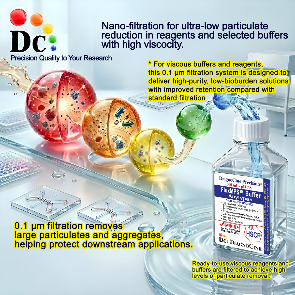
0.1 µm filtration removes large particulates and aggregates, helping protect downstream applications.
© Diagnocine® — DCP-GPBS1X
Where DCP-GPBS1X is used
A gelatin-based PBS blocking buffer for antibody-based detection assays.
Dot Blot & Protein Arrays
Blocks unoccupied binding sites on nitrocellulose and nylon membranes before antibody probing.
Wash, Dilution & Reconstitution
Stable pH 7.4 phosphate-buffered saline base suitable for wash and dilution steps in assay workflows.
iPSC-Derived Model Handling
Compatible with blocking and wash steps used in iPSC-derived model immunostaining workflows.
Endothelial & Primary Cell Models
Defined-pH buffer suited for wash steps with primary and endothelial cell models.
ELISA, Blotting & Blocking
Blocks non-specific binding sites on membranes and microplates, reducing background in Western blotting and ELISA.[1,2]
Microscopy & Optical Sensing
Low-particulate, defined-pH composition supports clean backgrounds in optical detection formats.
Specifications at a glance
All values below are as manufactured and quality-control tested for DCP-GPBS1X.
| Parameter | Specification |
|---|---|
| Formulation / Composition | PBS with 1% gelatin — see the full composition table below |
| Appearance | Clear, colorless liquid |
| pH USP <791> | 7.4 |
| Sodium Chloride Concentration | 137 mM |
| Potassium Chloride Concentration | 2.7 mM |
| Na2HPO4 (anhydrous) Concentration | 10.1 mM |
| KH2PO4 (anhydrous) Concentration | 1.76 mM |
| Gelatin Concentration | 1% |
| Parameter | Specification |
|---|---|
| Sterility | Nonsterile |
| DNase Activity | None detected (18 hr incubation with plasmid DNA at room temperature) |
| RNase Activity | None detected (18 hr incubation with ribosomal RNA at room temperature) |
| Filtration System | 0.1 µm membrane filtration (single step) |
| Water Quality | Ultrapure Type 1 water (18.2 MΩ·cm) USP <85> |
| Manufacturing Standard ISO 13485:2016 | Certified facilities |
| Parameter | Specification |
|---|---|
| Storage Temperature | 4°C (short-term); -20°C for long-term storage |
| Shelf Life | 6 months |
| Parameter | Specification |
|---|---|
| Manufacturing QMS | ISO 13485:2016-certified facility |
| Production Location | Final packaging, quality assurance, testing, and customization performed at DiagnoCine Precision, Totowa, New Jersey, USA |
| Intended Use | Research Use Only (RUO); not for clinical, diagnostic, or therapeutic use in humans |
| Membrane Compatibility | Compatible with nitrocellulose, nylon, and charge-modified nylon membranes; not recommended for PVDF membranes |
Full composition
Five named components at fixed concentrations, released per lot.
| Component | CAS Number | Concentration |
|---|---|---|
| Gelatin | 9000-70-8 | 1% |
| Sodium Chloride | 7647-14-5 | 137 mM |
| Potassium Chloride | 7447-40-7 | 2.7 mM |
| Sodium Phosphate Dibasic Anhydrous | 7558-79-4 | 10.1 mM |
| Potassium Phosphate Monobasic Anhydrous | 7778-77-0 | 1.76 mM |
Manufacturing & compliance
DCP-GPBS1X is manufactured, packaged, and tested under a controlled quality system.
ISO 13485:2016 QMS
Manufactured under ISO 13485-certified facilities (Suppliers of DiagnoCine Precision).
Ultrapure Type 1 Water
Formulated with Ultrapure Type 1 water (18.2 MΩ·cm) consistent with USP <85> expectations.
0.1 µm Filtration
0.1 µm filtration removes large particulates and aggregates, helping protect downstream applications.
Micro-Batch Precision
All final packaging, quality assurance, and testing are completed at the DiagnoCine R&D and Quality Testing Center, with specific customization requests assembled at DiagnoCine Precision in Totowa, New Jersey, USA.
Endotoxin
See CoA.
Particulate USP <788> Method 2
Particulate assessment follows USP <788> Method 2 as part of the filtration validation program.
Osmolality USP <785>
Osmolality testing is available within the USP <785> framework as part of the quality program.
Documentation / CoA
A Certificate of Analysis documenting appearance, pH, and DNase/RNase results is available on request.
How DCP-GPBS1X compares
DCP-GPBS1X against conventional gelatin blocking buffers.
| Parameter | DCP-GPBS1X | Conventional Buffer | Standard Alternative |
|---|---|---|---|
| DNase / RNase Validated | check_circle | cancel | cancel |
| Gelatin-Based Blocking Formulation | check_circle | cancel | cancel |
| Stable pH 7.4 Phosphate-Buffered Base | check_circle | check_circle | cancel |
| Water Quality | Ultrapure Type 1 (18.2 MΩ·cm) | Standard purified water | Standard purified water |
| Manufacturing QMS | ISO 13485:2016 | cancel | cancel |
| Membrane Compatibility Guidance | check_circle | cancel | cancel |
| Custom Formulation Available | check_circle | cancel | cancel |
Frequently asked questions
Common questions about DCP-GPBS1X.
Supporting literature
Curated references relevant to blocking buffer chemistry and immunoassay methodology.
- Kurien BT, Scofield RH. Western blotting. Methods. 2006;38(4):283-293. doi:10.1016/j.ymeth.2005.11.007
- Engvall E, Perlmann P. Enzyme-linked immunosorbent assay (ELISA). Immunochemistry. 1971;8(9):871-874. doi:10.1016/0019-2791(71)90454-X
- Mansfield MA. Rapid immunodetection on polyvinylidene difluoride membrane blots without blocking. Anal Biochem. 1995;229(1):140-143. doi:10.1006/abio.1995.1146
- Bass JJ, Wilkinson DJ, Rankin D, et al. An overview of technical considerations for Western blotting applications to physiological research. Scand J Med Sci Sports. 2017;27(1):4-25. doi:10.1111/sms.12702