

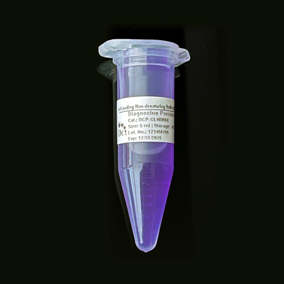
Gel Loading Non-denaturing Buffer [6X]
FluxMPS™ Gel Loading Non-denaturing Buffer [6X]
An MPS-grade, sterile 6X gel loading buffer engineered for DNA, RNA, and native protein sample preparation ahead of agarose or polyacrylamide gel electrophoresis. Built on a quadruple-stage filtration architecture (0.1 µm membrane filtration twice and 0.04 µm membrane filtration twice), it delivers ultra-clean, DNase- and RNase-free performance for demanding molecular biology and microfluidic sample-QC workflows.
- Quadruple-stage filtration: 0.1 µm membrane filtration twice and 0.04 µm membrane filtration twice
- Sterile, non-denaturing formulation compatible with DNA, RNA, and native protein samples
- Sucrose density agent (40%) ensures reliable sample sinking and sharp band resolution
- Bromophenol blue tracking dye for real-time visual monitoring of electrophoretic migration
- EDTA (0.1 M) inhibits nucleases and protects sample integrity
- No detectable DNase or RNase activity after 18 hr incubation testing
- pH 8.0, blue-purple appearance, 6X concentrate format
- Custom concentrations, pH, and additive formulations available on request
- Format6X Concentrate
- Size5 mL
- AppearanceBlue-purple
- pH8.0
- Filtration0.1 µm x2 + 0.04 µm x2
- SterilityFiltered in sterile environment
- DNase ActivityNone detected
- RNase ActivityNone detected
- Storage4°C (or -20°C long-term)
- Shelf Life6 months at 4°C
Engineered where standard loading buffers fall short
Conventional 0.22 µm-filtered gel loading buffers can carry subvisible particulates, inconsistent nuclease-free assurance, and undocumented purity claims that compromise sensitive downstream molecular and microfluidic workflows. FluxMPS™ buffers close that gap with documented, multi-stage filtration and lot-verified nuclease-free QC.
Microchannel-safe purity
Final 0.04 µm membrane filtration stage reduces particulate carryover relevant to USP <788> particulate control and fine-bore lab-on-a-chip channels.
Precise, stable pH
Formulated at pH 8.0, the non-denaturing chemistry preserves native DNA, RNA, and protein structure through loading and electrophoresis.
Ultrapure-grade water
Prepared with Ultrapure Type 1 water (18.2 MΩ·cm), consistent with USP <85> water-quality expectations for sensitive reagents.
Low background for imaging & documentation
Bromophenol blue tracking dye migrates cleanly alongside nucleic acids, giving a sharp, low-background visual reference under standard gel documentation systems.
Defined, traceable composition
Sucrose, EDTA, SDS, and bromophenol blue are each declared at their stated concentration — see the full composition table below.
Customization on demand
Alternate concentrations, additional chemicals, compounds, proteins, supplements, or a different pH can be formulated on request.
Quadruple-stage filtration system
Every lot is filtered through a sequential 0.1 µm and 0.04 µm membrane architecture, applied across four total passes, before sterile fill — positioning this reagent as an ultra-clean buffer for cell and molecular biology sample preparation.
-
1
0.1 µm Pre-filtration I
Removes large particulates and aggregates, extending the service life of downstream membranes.
-
2
0.04 µm Pre-filtration II
Retains fine particulates and bioburden below the size range of the smallest known mycoplasma types (about 0.2 microns), reducing mycoplasma-scale contamination risk.
-
3
0.1 µm Sterile-filtration I
A second 0.1 µm pass provides redundant particulate exclusion ahead of final polishing.
-
4
0.04 µm Sterile-filtration II — Final Polish
A second 0.04 µm pass completes the sterile filtration sequence within a controlled fill environment.
Performance vs. conventional buffer
Sequential 0.1 µm and 0.04 µm filtration, applied across four total passes, excludes finer particulates and mycoplasma-scale contaminants than a single 0.22 µm pass retains.

© Diagnocine® — DCP-GLNDB6X
Where DCP-GLNDB6X is used
A specialized reagent for preparing DNA, RNA, and native protein samples for loading onto agarose or polyacrylamide gels during electrophoresis, supporting restriction fragment analysis, PCR product analysis, and DNA ladder preparation.
Automated Bioreactors & Robotics
For laboratories running automated liquid-handling robots and high-throughput sample-loading platforms, an optional 0.01 µm (10 nm) ultra-filtered variant of this buffer is available to further reduce particulate load reaching precision dispensing hardware.
- Total Particulate Exclusion: finer sub-0.04 µm filtration for automated dispensing lines
- Valve & Sensor Protection: reduces particulate accumulation in liquid-handling robot valves and sensors
- Extended Reagent Stability: consistent formulation supports repeat automated dispensing cycles
Inquiry Required: the 0.01 µm (10 nm) ultra-filtered grade is available on request — contact support@diagnocine.com.
On-Chip Nucleic Acid Analysis
Compatible with lab-on-a-chip (LoC) electrophoretic separation modules used for compact DNA and RNA sizing within organ-on-a-chip (OoC) and tissue-on-a-chip (ToC) sample QC workflows.
Nucleic Acid & Protein Sample Loading
Mixed directly with DNA, RNA, or native protein samples before loading onto agarose or polyacrylamide gels, ensuring reliable well loading and dye-based migration tracking.
iPSC-Derived Model Genotyping
Supports gel-based verification of PCR amplicons, restriction digests, and vector constructs generated from iPSC-derived cell line workflows.
Genotyping & Expression Analysis
Enables gel electrophoresis-based confirmation of PCR amplicons and restriction digests from endothelial and primary cell genomic DNA extractions.
Native PAGE for Blotting Workflows
The non-denaturing formulation preserves native protein conformation, supporting blue native PAGE upstream of western blotting and immunodetection.
Migration Tracking & Optical Detection
Bromophenol blue tracking dye provides real-time visual monitoring of electrophoretic migration under standard gel documentation and optical detection systems.
Physical, chemical & quality parameters
All specifications below are drawn directly from lot-level quality control performed on DCP-GLNDB6X.
| Parameter | Specification |
|---|---|
| Formulation | Sucrose, EDTA, SDS, Bromophenol Blue (see composition table below) |
| Concentration | 6X Concentrate |
| Appearance | Blue-purple |
| pH USP <791> | 8.0 |
| Parameter | Specification |
|---|---|
| Filtration System | 0.1 µm membrane filtration twice and 0.04 µm membrane filtration twice |
| Sterility | Filtered in a sterile environment |
| DNase Activity | None detected (18 hr, plasmid DNA, room temperature) |
| RNase Activity | None detected (18 hr, ribosomal RNA, room temperature) |
| Water Quality | Ultrapure Type 1 water, 18.2 MΩ·cm |
| Manufacturing Standard | ISO 13485:2016 |
| Fill Environment | ISO Class 5 (Class 100) |
| Parameter | Specification |
|---|---|
| Storage Temperature | 4°C, or -20°C for longer-term storage |
| Shelf Life | 6 months at 4°C |
| Parameter | Specification |
|---|---|
| Manufacturing Facility | ISO 13485-certified, CE-approved supplier facilities (DiagnoCine Precision) |
| Quality Assurance & Testing | DiagnoCine R&D and Quality Testing Center |
| Custom Assembly Location | DiagnoCine Precision, Totowa, New Jersey, USA |
| Regulatory Alignment | CE-approved; ISO 13485:2016 aligned |
| Intended Use | For Research Use Only (RUO) |
Full composition
Every declared component of DCP-GLNDB6X, listed at its stated concentration.
| Component | CAS Number | Concentration |
|---|---|---|
| Sucrose | 57-50-1 | 40% |
| EDTA | 60-00-4 | 0.1 M |
| SDS | 151-21-3 | 0.5% |
| Bromophenol Blue | 115-39-9 | 0.05% |
Manufacturing & compliance
DCP-GLNDB6X is manufactured, filtered, and released under a documented quality system spanning supplier facilities and DiagnoCine's own testing center.
ISO 13485:2016 QMS
Manufactured under ISO 13485-certified and CE-approved supplier facilities.
Ultrapure Type 1 Water
Formulated with Ultrapure Type 1 water (18.2 MΩ·cm).
ISO Class 5 Fill & Finish
Final filtration and fill performed within an ISO Class 5 (Class 100) controlled environment.
Micro-Batch Precision
Final packaging, QA, and custom assembly are completed at DiagnoCine Precision in Totowa, New Jersey, USA.
DNase Activity (18 hr Assay)
None detected after incubation of plasmid DNA and this product for 18 hr at room temperature.
RNase Activity (18 hr Assay)
No RNase activity detected after incubation of ribosomal RNA and this product for 18 hr at room temperature.
pH Verification USP <791>
Each lot is verified to pH 8.0 prior to release.
Documentation & Certificate of Analysis
Certificate of Analysis available upon request, summarizing appearance, pH, filtration, sterility, and DNase/RNase QC results.
How DCP-GLNDB6X compares
A side-by-side view of the FluxMPS™ filtration architecture against conventional gel loading buffers.
| Parameter | DCP-GLNDB6X (FluxMPS™) | Conventional 0.22 µm-filtered buffer | Standard alternative (0.22 µm filtered buffer) |
|---|---|---|---|
| Final filtration pore size | 0.04 µm | 0.22 µm | 0.22 µm |
| Number of filtration passes | 4 | 1 | 1 |
| DNase/RNase activity tested | check_circle | cancel | cancel |
| Non-denaturing formulation | check_circle | check_circle | check_circle |
| Bromophenol blue tracking dye included | check_circle | check_circle | cancel |
| Manufacturing QMS (ISO 13485:2016) | check_circle | cancel | cancel |
| Ultrapure Type 1 water (18.2 MΩ·cm) | check_circle | cancel | cancel |
| Custom formulation available | check_circle | cancel | cancel |
Frequently asked questions
Answers to common questions about DCP-GLNDB6X.
Supporting literature
Curated references relevant to gel loading buffer chemistry and nucleic acid electrophoresis.
- Lee, P.Y., Costumbrado, J., Hsu, C.Y., Kim, Y.H. Agarose gel electrophoresis for the separation of DNA fragments. J Vis Exp. 2012. doi:10.3791/3923
- Voytas, D. Agarose gel electrophoresis. Curr Protoc Mol Biol. 2001. doi:10.1002/0471142727.mb0207s51
- Green, M.R., Sambrook, J. Analysis of DNA by agarose gel electrophoresis. Cold Spring Harb Protoc. 2019. doi:10.1101/pdb.top100388
- Ornstein, L. Disc electrophoresis I: background and theory. Ann N Y Acad Sci. 1964. doi:10.1111/j.1749-6632.1964.tb14847.x
- Laemmli, U.K. Cleavage of structural proteins during the assembly of the head of bacteriophage T4. Nature. 1970. doi:10.1038/227680a0
- Huang, Y. et al. Microfluidic capillary electrophoresis chips. Electrophoresis. 2013. doi:10.1002/elps.201200574
- Kricka, L.J. Miniaturization of analytical systems. Clin Chem. 1998. doi:10.1093/clinchem/44.9.2008
- Karger, B.L., Chu, Y.H., Foret, F. Capillary electrophoresis of proteins and nucleic acids. Anal Chem. 2000. doi:10.1021/ac000041j