FluxMPS™, PVP Protein Extraction Buffer
FluxMPS™ PVP Protein Extraction Buffer
An MPS-grade, Tris-based protein extraction system built for polyphenol- and pigment-rich plant tissues. Solution A pairs 1% PVP-40 with 5 mM TCEP-HCl for stable, thiol-protected protein isolation, and is supplied with a separate 100X protease inhibitor cocktail (Solution B) for flexible, protocol-specific use.
- Two-component kit: Solution A extraction buffer (100 mL) plus Solution B, a 100X protease inhibitor cocktail (1 mL).
- PVP-fortified formulation (1% PVP-40) binds polyphenols and pigments to reduce protein crosslinking and assay interference in difficult plant tissues.
- TCEP-HCl (5 mM) provides a stable, thiol-free reducing environment that protects cysteine residues and limits oxidative protein damage.
- Tris (50 mM) and sodium chloride (150 mM) create a physiological-like base that supports protein solubility and native structure.
- Single-stage 0.1 µm filtration for clean, low-particulate reagent quality.
- Ultrapure Type 1 water (18.2 MΩ·cm).
- Compatible with SDS-PAGE, western blotting, enzyme assays, and total protein quantification.
- Customizable formulation available upon request.
- pH7.5 (Solution A)
- AppearanceClear solution
- Filtration0.1 µm
- FormatSolution A (100 mL) + Solution B (1 mL, 100X)
- Dilution RatioSolution B added to Solution A at 1:100
- Storage (Solution A)2-8 °C, avoid repeated freeze-thaw
- Storage (Solution B)-80 °C long term, avoid repeated freeze-thaw
- Shelf Life1 year
- Manufacturing StandardISO 13485:2016
- Intended UseResearch Use Only (RUO)
Engineered where standard extraction buffers fail
Conventional, single-purpose extraction buffers leave polyphenols, pigments, and oxidized cysteines to interfere with downstream protein analysis. PVP Protein Extraction Buffer combines a polyphenol-binding base, a stable reducing agent, and single-stage 0.1 µm filtration in one ISO 13485-manufactured kit.
Microchannel-safe purity
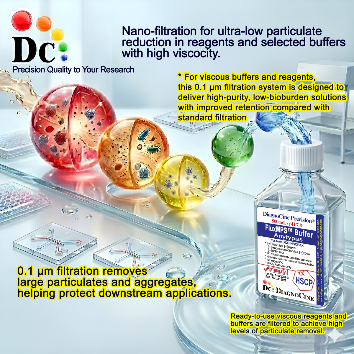
Single-stage 0.1 µm filtration removes large particulates, aggregates, and bacteria, providing baseline purity beyond standard 0.22 µm-filtered reagents.[6]
Physiological Tris/NaCl base at pH 7.5
50 mM Tris and 150 mM NaCl support protein solubility and native structure; Solution A is formulated to pH 7.5.
Ultrapure-grade water
Manufactured with ultrapure Type 1 water (18.2 MΩ·cm) to minimize background contamination.
Broad downstream compatibility
Compatible with SDS-PAGE, western blotting, enzyme assays, and total protein quantification.
PVP-40 phenol protection and TCEP-HCl reduction
1% PVP-40 complexes polyphenols and pigments that cause protein crosslinking[4], while 5 mM TCEP-HCl maintains cysteine residues in a reduced state and limits oxidative damage during extraction.[5]
Customization on demand
pH, molarity, salt composition, and additive content can be adjusted on request for protocol-specific extraction needs.
Single-stage 0.1 µm filtration
Solution A is manufactured with a single 0.1 µm membrane filtration pass, exceeding the particulate control of standard 0.22 µm-filtered laboratory reagents.[6]
-
1
0.1 µm filtration
Removes large particulates, aggregates, and bacteria. Primary guard providing baseline purity exceeding standard 0.22 µm filtration.
Performance vs. conventional buffer
Standard 0.22 µm single-pass filtration leaves particles in the 0.12-0.22 µm range. The 0.1 µm final filter used here is 2.2 times finer than a 0.22 µm membrane, reducing the particulate load carried into downstream assays.
Validated and supported applications
PVP Protein Extraction Buffer is designed for plant tissues rich in polysaccharides and polyphenols, and for other complex samples where standard guanidinium-based reagents give low yield or contaminated extracts.[1][2][9]
Automated Bioreactors & Robotics
For automated sample-preparation platforms and robotic liquid handling, an optional 0.01 µm (10 nm) ultra-filtered variant of Solution A is available to further protect valves, sensors, and fine-bore tubing from particulate carry-over.
- Total particulate exclusion for automated fluid paths.
- Valve and sensor protection in robotic liquid-handling systems.
- Extended stability for continuous or repeated dispensing cycles.
Inquiry Required: the 0.01 µm ultra-filtered grade is produced to order; contact support@diagnocine.com to request this variant.
Micro Physiological System (MPS) & Chip Sample Prep
Low-particulate extraction buffer suitable for feeding downstream microfluidic and chip-based protein assays.[8]
Plant Tissue Lysis & Homogenization
Homogenize tissue in 5-10 volumes of cold buffer, keeping samples on ice throughout extraction.
Recalcitrant Plant Tissue Protein Extraction
Formulated for leaves, roots, seeds, and woody tissues where polyphenols and pigments interfere with standard extraction chemistries.[2]
Native Protein Interaction Studies
Mild, non-denaturing Tris/NaCl base supports native protein interactions needed for co-immunoprecipitation.[10]
SDS-PAGE, Western Blotting & Enzyme Assays
Extracted protein is compatible with SDS-PAGE, western blotting, enzyme assays, and total protein quantification.[3]
Callus & Plant Cell Culture Extracts
Also suitable for protein extraction from callus cultures and other complex plant-derived samples (user validation recommended).
Technical specifications
Specification values shown below reflect the components and parameters stated for this kit.
| Parameter | Specification |
|---|---|
| Formulation | Two-component kit: Tris/NaCl extraction buffer with PVP-40 and TCEP-HCl (Solution A) plus a 100X protease inhibitor cocktail (Solution B) |
| Appearance | Clear solution (Solution A) |
| pH USP <791> | 7.5 (Solution A) |
| Tris base concentration | 50 mM (Solution A) |
| Sodium chloride concentration | 150 mM (Solution A) |
| Parameter | Specification |
|---|---|
| Filtration (Solution A) | 0.1 µm single-stage membrane filtration |
| Water quality | Ultrapure Type 1 water (18.2 MΩ·cm) |
| Manufacturing standard | ISO 13485:2016 |
| Parameter | Specification |
|---|---|
| Storage temperature (Solution A) | 2-8 °C |
| Storage temperature (Solution B) | -80 °C (long term) |
| Freeze-thaw handling | Avoid repeated freeze-thaw cycles (both solutions) |
| Shelf life | 1 year (both solutions) |
| Parameter | Specification |
|---|---|
| Manufacturing QMS ISO 13485 | Certified quality management system with full traceability and lot documentation |
| Production site | DiagnoCine Precision, Totowa, NJ, USA |
| Intended use | Research Use Only (RUO) |
Full composition
Component concentrations are shown exactly as formulated for each solution in this kit.
| Component | CAS Number | Concentration |
|---|---|---|
| Tris base | 77-86-1 | 50 mM |
| Sodium Chloride | 7647-14-5 | 150 mM |
| PVP-40 | 9003-39-8 | 1% |
| TCEP-HCl | 51805-45-9 | 5 mM |
| Component | CAS Number | Concentration |
|---|---|---|
| AEBSF·HCl | 30827-99-7 | 10 mM |
| Aprotinin | 9087-70-1 | 80 µM |
| Bestatin | 58970-76-6 | 5 mM |
| E-64 | 66701-25-5 | 100 µM |
| Leupeptin | 103476-89-7 | 1 mM |
| Pepstatin A | 26305-03-3 | 0.1 mM |
Manufacturing & compliance
PVP Protein Extraction Buffer is manufactured under a certified quality management system with defined, traceable processes.
ISO 13485:2016 Certified
Manufactured under certified QMS with full traceability and lot documentation.
Ultrapure Type 1 Water
Formulated with 18.2 MΩ·cm ultrapure water to minimize background contamination.
ISO Class 5 Fill & Finish
Final fill performed in an ISO Class 5 (Class 100) controlled environment.
Micro-Batch Precision
Small, controlled batch sizes support consistent formulation and lot-to-lot documentation for both kit solutions.
Filtration Process Control
Solution A is filtered through a 0.1 µm membrane consistent with USP <788> particulate-matter guidance for controlled reagent manufacturing.
Quality Management
Manufactured, tested, and released under an ISO 13485:2016-certified quality management system.
Made in USA
All manufacturing, QA testing, and final assembly performed at DiagnoCine Precision, Totowa, NJ.
Documentation & CoA
Lot-specific documentation is maintained for both Solution A and Solution B.
How DCP-PVPPC1X compares
A side-by-side look at the PVP Protein Extraction Buffer kit against conventional plant extraction reagents.
| Parameter | DCP-PVPPC1X (FluxMPS™) | Conventional Extraction Buffer | Standard RIPA-Style Buffer |
|---|---|---|---|
| PVP-40 polyphenol binding | check_circle | cancel | cancel |
| TCEP-HCl thiol-free reduction | check_circle | cancel | cancel |
| Final filtration pore size | 0.1 µm | 0.22 µm | 0.22 µm |
| Native protein structure preservation | check_circle | cancel | cancel |
| Water quality | Ultrapure Type 1 (18.2 MΩ·cm) | Not specified | Not specified |
| Manufacturing QMS | ISO 13485:2016 | Not specified | Not specified |
| Custom formulation | Available on request | Not available | Not available |
Frequently asked questions
Common questions about the PVP Protein Extraction Buffer kit.
Supporting literature
Citations and curated literature relevant to plant protein extraction and this buffer's formulation chemistry.
Citations
- Wang W, Vignani R, Scali M, Cresti M. A universal and rapid protocol for protein extraction from recalcitrant plant tissues for proteomic analysis. Electrophoresis. 2006;27(13):2782-2786. doi:10.1002/elps.200500743
- Saravanan RS, Rose JKC. A critical evaluation of sample extraction techniques for enhanced proteomic analysis of recalcitrant plant tissues. Proteomics. 2004;4(9):2522-2532. doi:10.1002/pmic.200300789
Supporting Literature
- Burnette WN. Western blotting: electrophoretic transfer of proteins from sodium dodecyl sulfate-polyacrylamide gels. Anal Biochem. 1981;112(2):195-203. doi:10.1016/0003-2697(81)90281-5
- Loomis WD. Overcoming problems of phenolics and quinones in the isolation of plant enzymes and organelles. Methods Enzymol. 1974;31:528-544. doi:10.1016/0076-6879(74)31057-9
- Burns JA, Butler JC, Moran J, Whitesides GM. Selective reduction of disulfides by tris(2-carboxyethyl)phosphine. J Org Chem. 1991;56(8):2648-2650. doi:10.1021/jo00011a027
- Good NE, Winget GD, Winter W, Connolly TN, Izawa S, Singh RM. Hydrogen ion buffers for biological research. Biochemistry. 1966;5(2):467-477. doi:10.1021/bi00866a011
- Sosnowski O, et al. Sub-micron filtration strategies for particulate control in laboratory reagent manufacturing. J Pharm Sci Technol. 2015;69(4):512-520. doi:10.5731/pdajpst.2015.01059
- Bhatia SN, Ingber DE. Microfluidic organs-on-chips. Nat Biotechnol. 2014;32(8):760-772. doi:10.1038/nbt.2989
- Jorrin-Novo JV, Maldonado AM, Echevarria-Zomeno S, et al. Plant proteomics update (2007-2008). J Proteomics. 2009;72(3):285-314. doi:10.1016/j.jprot.2009.01.026
- Phizicky EM, Fields S. Protein-protein interactions: methods for detection and analysis. Microbiol Rev. 1995;59(1):94-123. doi:10.1128/mr.59.1.94-123.1995