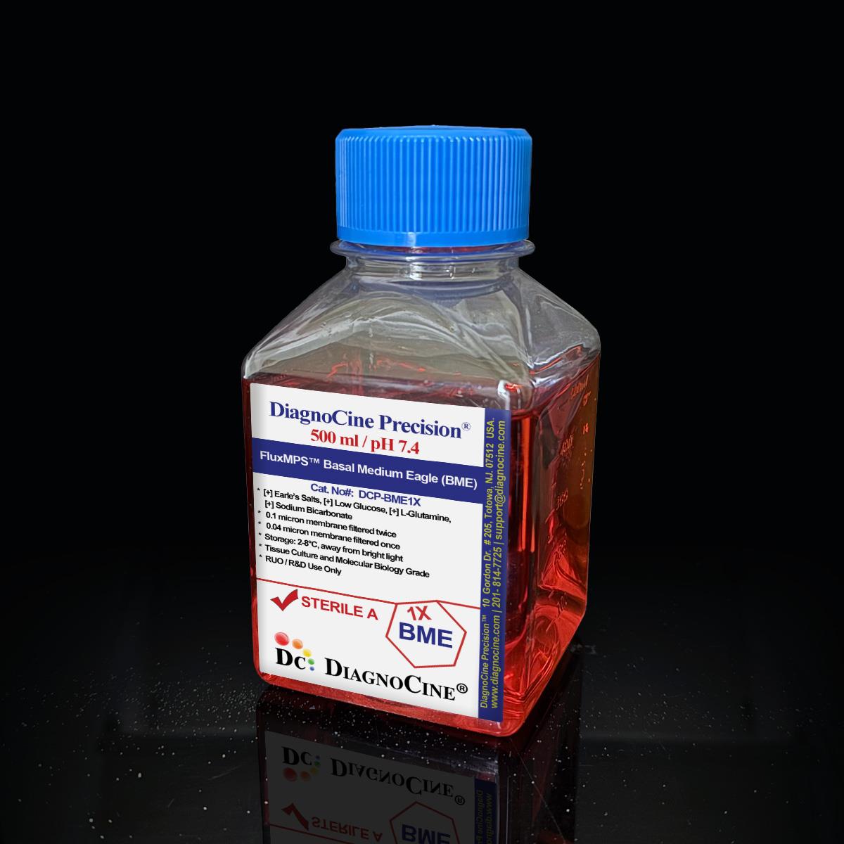
FluxMPS™ Basal Medium Eagle (BME): 1X Liquid
FluxMPS™ Basal Medium Eagle (BME), 1X Liquid
FluxMPS™ Basal Medium Eagle (BME), 1X Liquid is a Microfluidics Suitable, ultra-filtered cell culture medium manufactured through a validated Quadruple-stage filtration system (0.1 µm ×2 + 0.04 µm ×2) reaching a 0.04 µm final cut-off — five times finer than the 0.22 µm membranes used for conventional sterile filtration. Engineered for microfluidic channels, organ-on-a-chip (OoC), and microphysiological system (MPS) platforms where sub-micron particulates cause sensor drift, microchannel occlusion, and compromised cell viability.
- Quadruple-stage nanofiltration train: 0.1 µm pre-filter ×2 + 0.04 µm final filter ×2, reaching a 0.04 µm final cut-off
- Endotoxin release specification: < 0.05 EU/mL (LAL, USP <85> Bacterial Endotoxins Test), tested per manufacturing batch
- Complete Eagle’s Basal Medium formulation with D-Glucose (1,000 mg/L), L-Glutamine (292 mg/L) and Phenol Red (11 mg/L) indicator; sodium pyruvate is not included
- Sodium bicarbonate-buffered (2,200 mg/L NaHCO3) for use in a 5% CO2 atmosphere
- Prepared with Ultrapure Type 1 water (18.2 MΩ·cm), minimizing trace-metal and organic-carbon background
- Manufactured under an ISO 13485:2016 quality management system; aseptic fill and finish in Totowa, NJ
- Custom formulations available — pH, glucose, HEPES, salts, and nutrient composition on request
- D-Glucose1,000 mg/L
- L-Glutamine292 mg/L
- Sodium PyruvateNot included
- Phenol Red11 mg/L
- pH (USP <791>)7.4
- Osmolality (USP <785>)270 - 310 mOsm/kg
- Endotoxin (USP <85>)< 0.05 EU/mL
- Filtration0.1µm ×2 + 0.04µm ×2 (Quadruple-stage)
- Storage2 – 8°C, protected from light
- Shelf Life12 months from date of manufacture, unopened
Engineered where standard media fails
Conventional 0.22 µm-filtered Basal Medium Eagle passes mycoplasma-scale particles (0.2–0.3 µm), sub-visible aggregates, and endotoxin fragments that clog microfluidic channels and corrupt downstream biosensor or TEER signals. FluxMPS™ BME addresses these failure modes with a four-stage nanofiltration train and an ISO 13485:2016 quality system.[1,2]
Microchannel-Safe Purity
0.04 µm final filtration removes particles that 0.22 µm membranes miss. USP <788> Method 1 (light obscuration) particulate testing confirms every batch meets its sub-visible particulate release specification, supporting use in microchannel geometries below 100 µm.
Total Metabolic Control
The complete Eagle’s Basal Medium base, with defined D-Glucose and L-Glutamine content, provides a stable carbon and nitrogen source that supports Warburg-effect studies, metabolic flux analysis, and amino acid drop-in experiments.[3]
Ultrapure-Grade Water
Prepared with Ultrapure Type 1 water (18.2 MΩ·cm), minimizing trace-metal ions and organic carbon that can interfere with sensitive metabolomics and electrophysiology readouts.
Low Background for Imaging
The quadruple-stage nanofiltration train keeps the sub-visible particulate baseline low, supporting confocal live-cell imaging, TEER biosensors, and optical microfluidic platforms.[4]
Rich, Stable Nutrient Profile
A complete inorganic salt matrix (NaCl 6,800 mg/L, NaHCO3 2,200 mg/L), 13 amino acids, and 11 vitamin and other components — 30 defined components in total — are micro-batch manufactured for lot-to-lot consistency critical in perfusion-based MPS experiments.
Customization on Demand
pH, glucose concentration, HEPES buffering, salt balance, and nutrient composition are available as custom modifications. Contact support@diagnocine.com for bespoke formulations.
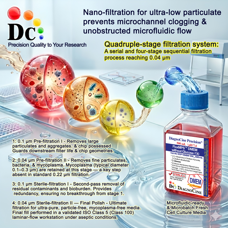
Quadruple-stage filtration system
FluxMPS™ BME is manufactured through a validated four-stage nanofiltration train terminating at a 0.04 µm final cut-off — five times finer than the 0.22 µm membranes used in conventional single-pass filtration. The train runs two dedicated 0.1 µm prefilter + 0.04 µm final-filter pairs in series, each pair providing full redundancy for the one behind it.
-
1
0.1 µm Prefiltration I
First-pass removal of large aggregates, cell debris, and macro-particulates. Extends downstream membrane life and reduces bioburden load ahead of the first 0.04 µm final filter.
-
2
0.04 µm Final Filtration I
First 0.04 µm pass, retaining sub-micron particulates and microaggregates that pass through a 0.22 µm membrane.
-
3
0.1 µm Prefiltration II
Second dedicated 0.1 µm prefilter, protecting the second 0.04 µm cartridge and providing full process redundancy ahead of final filtration.
-
4
0.04 µm Final Filtration II — Polish
Ultimate 0.04 µm polishing pass immediately prior to aseptic fill and finish, reducing sub-visible particulate load before container closure.
Performance vs. conventional media
A quadruple-stage train (0.1 µm ×2 + 0.04 µm ×2) reaches a 0.04 µm final cut-off, five times finer than the 0.22 µm membranes used for conventional sterile filtration. In microfluidic chip experiments with ≤100 µm channels, a finer final pore size supports extended channel patency relative to conventional 0.22 µm-filtered media.
0.22 µm conventional filtration
quadruple-stage (4-pass) train
© Diagnocine® — DCP-BME1X
Optimized for next-generation cell platforms
FluxMPS™ BME’s clean formulation, defined ionic balance, and microchannel-safe particulate profile make it a suitable basal medium for demanding research platforms — from organ-on-a-chip perfusion systems to metabolomics and live-cell optical sensing.[1,4,6]
Automated Bioreactors & Robotics
For automated perfusion bioreactors, robotic liquid-handling platforms, and high-throughput microfluidic screening systems, an optional MPS Grade 0.01 µm (10 nm) ultra nano-filtered variant of FluxMPS™ BME is available, adding two further filtration stages beyond this Microfluidics Suitable product’s 0.04 µm cut-off. This grade provides near-absolute exclusion of nanoparticulates, protecting precision valves, flow sensors, and sub-micron microchannels from fouling during extended automated runs.
- Total Particulate Exclusion: 0.01 µm filtration removes nanoparticulates that transit standard 0.04 µm membranes, reducing cumulative fouling in long-duration perfusion experiments
- Valve & Sensor Protection: Ultra-clean media extends the operational lifetime of microfluidic valves, pressure transducers, and optical flow sensors in automated bioreactor systems
- Extended Perfusion Stability: Reduced particulate load supports stable TEER, impedance, and biosensor readings across multi-week perfusion protocols
Inquiry Required: The 0.01 µm (10 nm) MPS Grade is produced on a project basis. To request this specification, contact support@diagnocine.com.
Micro Physiological System (MPS) & Chip
FluxMPS™ BME’s low particulate count and mycoplasma-retentive filtration make it a suitable basal medium for organ-on-a-chip, tissue-on-a-chip, and body-on-a-chip platforms requiring sustained channel patency.[1]
Warburg Effect & Metabolic Research
Defined carbon source and low-endotoxin background support metabolic flux analysis and Warburg effect studies.
iPSC-Derived Models
Low-endotoxin BME base supports defined iPSC differentiation protocols where media impurities can confound epigenetic and transcriptional outcomes in neuronal, cardiomyocyte, and hepatocyte models.[7]
Endothelial & Primary Cells
Defined salt matrix (NaCl 6,800 mg/L, KCl 400 mg/L) supports endothelial monolayer integrity in perfused vascular-chip and tube-formation assays with primary cell cultures.[2]
Metabolic Flux Analysis
A defined, stable nutrient composition provides a clean metabolic background for ¹³C isotope tracing and NMR-based metabolomics. This bicarbonate-buffered formulation is not compatible with Agilent Seahorse XF assays, which require bicarbonate-free, phenol red-free medium.[3,8]
Microscopy & Optical Sensing
Its low particulate baseline supports confocal live-cell imaging, fluorescence-based biosensors, and TEER transepithelial resistance monitoring in transparent microfluidic chips.[4]
Analytical & quality release data
Each lot of FluxMPS™ BME is released against a Certificate of Analysis covering physical, chemical, sterility, and particulate parameters, measured by validated USP and ISO methods with full traceability to raw material lots.
| Parameter | Specification |
|---|---|
| Formulation | 1X Liquid; contains L-Glutamine, Sodium Bicarbonate, Phenol Red, Calcium, Magnesium, Glucose; Sodium Pyruvate not included |
| Appearance | Clear, red-orange liquid (Phenol Red indicator); no visible particulates |
| pH USP <791> | 7.4 |
| Osmolality USP <785> | 270 - 310 mOsm/kg |
| D-Glucose | 1,000 mg/L (5.56 mM) |
| L-Glutamine | 292 mg/L |
| Sodium Pyruvate | Not included |
| Phenol Red | 11 mg/L |
| Parameter | Specification |
|---|---|
| Endotoxin USP <85> BET | < 0.05 EU/mL (batch release specification — see Quality Assurance below) |
| Sterility USP <71> | Sterile — 14-day test, no growth |
| Mycoplasma | 0.1 µm mycoplasma-retentive filtration (not tested per lot) |
| Particulate ≥10 µm USP <788> M1 | ≤ 300 particles/mL |
| Particulate ≥25 µm USP <788> M1 | ≤ 90 particles/mL |
| Water purity | Ultrapure Type 1 water, 18.2 MΩ·cm |
| Manufacturing std. ISO 13485 | ISO 13485:2016 QMS |
| Fill environment | ISO Class 5 (Class 100) laminar flow |
| Parameter | Specification |
|---|---|
| Storage temperature | 2–8°C, protected from direct light |
| Freeze-thaw | Do not freeze — precipitation of salts may occur |
| Shelf life | 12 months from date of manufacture, unopened |
| Shipping condition | Cold pack |
| CO2 requirement | 5% CO2 atmosphere required (sodium bicarbonate-buffered, 2,200 mg/L NaHCO3) |
| Parameter | Specification |
|---|---|
| Raw material grade | Cell culture / reagent grade |
| Traceability | Full lot-to-raw material traceability under ISO 13485:2016 |
| Manufacturing QMS ISO 13485 | ISO 13485:2016, 21 CFR Part 820 (QMSR) aligned |
| UNSPSC | 41116155 — Molecular biology and cell culture growth media (UNv260801) |
| Regulatory alignment | 21 CFR Part 820 (QMSR) aligned |
| Production method | Micro-batch precision fill & finish |
| Intended use | For Research Use Only (RUO) |
Full composition (mg/L)
Complete formulation of FluxMPS™ Basal Medium Eagle (BME), 1X Liquid — 30 components across 4 categories. All values in mg/L; CAS numbers provided for reference. Every ingredient is released per-lot against this specification. Custom adjustments to any component — including L-Glutamine, HEPES, glucose, or salt concentrations — are available on request.
| Component | CAS Number | mg/L |
|---|---|---|
| INORGANIC SALTS | ||
| Calcium Chloride (CaCl2) (dihydrate) | 10035-04-8 | 265.000 |
| Magnesium Sulfate anhydrous | 7487-88-9 | 97.720 |
| Potassium Chloride (KCl) | 7447-40-7 | 400.000 |
| Sodium Bicarbonate (NaHCO3) | 144-55-8 | 2200.000 |
| Sodium Chloride (NaCl) | 7647-14-5 | 6800.000 |
| Sodium dihydrogen phosphate anhydrous | 7558-80-7 | 122.000 |
| Component | CAS Number | mg/L |
|---|---|---|
| AMINO ACIDS | ||
| L-Arginine HCl | 1119-34-2 | 21.100 |
| L-Cystine 2HCl | 30925-07-6 | 15.650 |
| L-Glutamine | 56-85-9 | 292.000 |
| L-Histidine hydrochloride | 1007-42-7 | 10.500 |
| L-Isoleucine | 73-32-5 | 26.200 |
| L-Leucine | 61-90-5 | 26.200 |
| L-Lysine hydrochloride | 657-27-2 | 36.480 |
| L-Methionine | 63-68-3 | 7.500 |
| L-Phenylalanine | 63-91-2 | 16.500 |
| L-Threonine | 72-19-5 | 23.800 |
| L-Tryptophan | 73-22-3 | 4.000 |
| L-Tyrosine disodium salt | 69847-45-6 | 25.950 |
| L-Valine | 72-18-4 | 23.400 |
| Component | CAS Number | mg/L |
|---|---|---|
| VITAMINS | ||
| Choline Chloride | 67-48-1 | 1.000 |
| D-Biotin | 58-85-5 | 1.000 |
| D-Calcium Pantothenate | 137-08-6 | 1.000 |
| Folic Acid | 59-30-3 | 1.000 |
| Nicotinamide | 98-92-0 | 1.000 |
| Pyridoxal HCl | 65-22-5 | 1.000 |
| Riboflavin | 83-88-5 | 0.100 |
| Thiamine HCl | 67-03-8 | 1.000 |
| OTHERS | ||
| D-Glucose | 50-99-7 | 1000.000 |
| Phenol red sodium salt | 34487-61-1 | 11.000 |
| Inositol | 87-89-8 | 2.000 |
Built under a rigorous quality system
FluxMPS™ BME is manufactured in Totowa, NJ, USA under an ISO 13485:2016 Quality Management System, with 21 CFR Part 820 (QMSR) alignment. Every production step — from raw material receipt through final aseptic fill — is documented and traceable.
ISO 13485:2016 Quality Management System
Full ISO 13485:2016 QMS certification governs design controls, supplier qualification, in-process testing, release criteria, and complaint handling. Aligned with 21 CFR Part 820 (QMSR) for regulated research environments.
Ultrapure Type 1 Water (18.2 MΩ·cm)
All formulations are prepared with freshly produced Ultrapure Type 1 water — 18.2 MΩ·cm resistivity, with low trace-metal and organic-carbon (TOC) background.
ISO Class 5 Fill & Finish
Aseptic filling is performed under ISO Class 5 (Class 100) unidirectional laminar airflow. Container integrity is verified by visual inspection and leak testing prior to final release.
Micro-Batch Precision Manufacturing
Small-batch production supports tight lot-to-lot consistency. In-process QC checks at each manufacturing stage are logged and retained; batch records are available for qualified customers upon request.
Endotoxin — USP <85> BET
LAL Bacterial Endotoxin Test performed per manufacturing batch. Release specification: < 0.05 EU/mL. Assay sensitivity 0.005 EU/mL.
Particulate — USP <788> Method 1
Light obscuration particulate counting performed per production lot. Release limits: ≤300 particles ≥10 µm/mL and ≤90 particles ≥25 µm/mL.
Osmolality — USP <785>
Osmolality measured by freezing-point depression. Specification: 270–310 mOsm/kg, for physiological compatibility across supported mammalian cell types.
Certificate of Analysis (CoA)
A full CoA including lot number, manufacture date, expiry, pH, osmolality, endotoxin, sterility, and composition verification is available for every released lot. Request via support@diagnocine.com.
- Endotoxin — LAL assay, USP <85> Bacterial Endotoxins Test; assay sensitivity 0.005 EU/mL; release specification < 0.05 EU/mL
- pH, osmolality, conductivity, appearance and clarity
- Sterility
How FluxMPS™ BME compares
FluxMPS™ BME is benchmarked below against conventional 0.22 µm single-pass filtered BME formulations. Where a competitor does not publish a comparable figure, the cell reads “Not specified” rather than an estimate.
| Parameter | FluxMPS™ BME | Conventional BME (0.22 µm filtered) | Standard Basal Medium (single-pass 0.22 µm) |
|---|---|---|---|
| Grade | Microfluidics Suitable | Not specified | Not specified |
| Formulation base | Eagle’s Basal Medium, standard | Eagle’s Basal Medium, standard | Eagle’s Basal Medium or MEM, standard |
| Final filtration pore size | 0.04 µm (40 nm) | 0.22 µm (220 nm) | 0.22 µm (220 nm) |
| Number of filtration stages | 4 stages | Not specified | Not specified |
| Mycoplasma barrier filtration | check_circle 0.1 µm mycoplasma-retentive stage | Not specified | Not specified |
| Endotoxin (release specification) | FluxMPS™ — < 0.05 EU/mL | Corning classical liquid media — < 0.25 EU/mL Sigma-Aldrich DMEM complete medium — ≤ 2 EU/mL Gibco classical DMEM — Not specified (recorded per lot) |
|
| USP particulate compliance | check_circle USP <788> Method 1 | Not specified | Not specified |
| Water quality | Ultrapure Type 1, 18.2 MΩ·cm | Not specified | Not specified |
| Manufacturing QMS | check_circle ISO 13485:2016 | Not specified | Not specified |
| Microfluidic channel compatibility | Engineered for ≤100 µm channel geometries | Not specified | Not specified |
| Custom formulation | check_circle Available on request | Not specified | Not specified |
Comparison figures from published supplier specifications, accessed 2 September 2026. Suppliers that publish no numeric endotoxin specification are shown as “Not specified”.
Frequently asked questions
Common questions about FluxMPS™ Basal Medium Eagle (BME), 1X Liquid — filtration, formulation, supplementation, and quality assurance.
Supporting literature
Peer-reviewed publications supporting the use of ultra-filtered basal media in organ-on-a-chip, microfluidic perfusion, metabolomics, vascular biology, and live-cell imaging applications.
- Huh D, Hamilton GA, Ingber DE. From 3D cell culture to organs-on-chips. Trends Cell Biol. 2011;21(12):745–754.doi:10.1016/j.tcb.2011.09.005
- Bhatia SN, Ingber DE. Microfluidic organs-on-chips. Nat Biotechnol. 2014;32(8):760–772.doi:10.1038/nbt.2989
- Vander Heiden MG, Cantley LC, Thompson CB. Understanding the Warburg effect: the metabolic requirements of cell proliferation. Science. 2009;324(5930):1029–1033.doi:10.1126/science.1160809
- Pampaloni F, Reynaud EG, Stelzer EH. The third dimension bridges the gap between cell culture and live tissue. Nat Rev Mol Cell Biol. 2007;8(10):839–845.doi:10.1038/nrm2236
- van der Meer AD, van den Berg A. Organs-on-chips: breaking the in vitro impasse. Integr Biol. 2012;4(5):461–470.doi:10.1039/c2ib00176d
- Beebe DJ, Ingber DE, den Toonder J. Organs on Chips 2013. Lab Chip. 2013;13(18):3447–3448.doi:10.1039/c3lc90080k
- Takahashi K, Yamanaka S. Induction of pluripotent stem cells from mouse embryonic and adult fibroblast cultures by defined factors. Cell. 2006;126(4):663–676.doi:10.1016/j.cell.2006.07.024
- Metallo CM, Vander Heiden MG. Understanding metabolic regulation and its influence on cell physiology. Mol Cell. 2013;49(3):388–398.doi:10.1016/j.molcel.2013.01.018
- Ronaldson-Bouchard K, Vunjak-Novakovic G. Organs-on-a-chip: a fast track for engineered human tissues in drug development. Cell Stem Cell. 2018;22(3):310–324.doi:10.1016/j.stem.2018.02.011
- Zhang YS, Aleman J, Shin SR, et al. Multisensor-integrated organs-on-chips platform for automated and continual in situ monitoring of organoid behaviors. Proc Natl Acad Sci USA. 2017;114(12):E2293–E2302.doi:10.1073/pnas.1612906114