


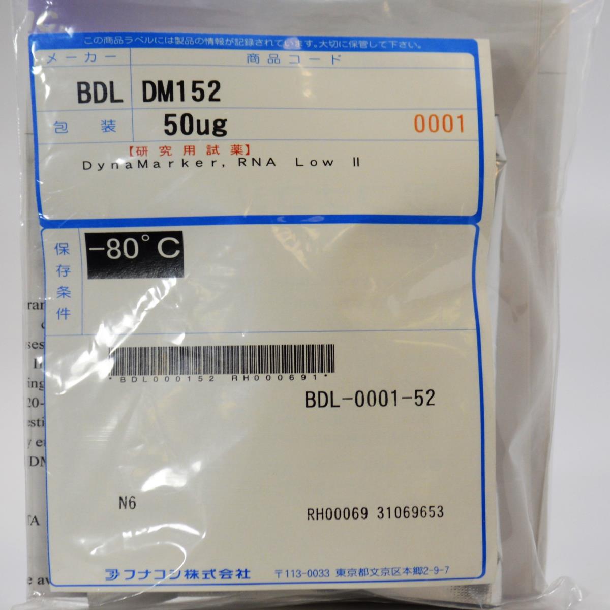
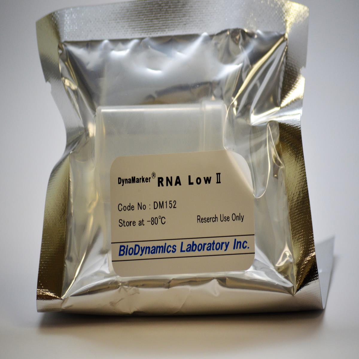
DynaMarker, RNA Low II
DynaMarker, RNA Low II
Cat. No. FNK-DM152
Size 50 μg (72 μl), 0.7 mg/ml
Storage -80°C
Range 20-500 base of RNA
Description
This product is research use only
The DynaMarker RNA Low II consists of seven single-stranded RNAs. The 20-base and 50-base RNA are synthesized by chemically (not phosphorylated). The 100, 200, 300, 400 and 500 bases are synthesized by in vitro transcription. The DynaMarker RNA Low II is suitable for determinating size of single-stranded RNAs in denaturing polyacrylamide gel electrophoresis. The concentration of each RNA (20-500 base) in the marker is approximately 0.1 μg/μl. It is useful for estimating of RNA amount. The DynaMarker RNA Low II can be visualized by ethidium bromide staining or by Gel IndicatorTM RNA Staining Solution (DM590, 595).
DynaMarker RNA Low II consists of seven single-stranded RNAs(20-500bases)
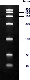
Electrophoresis profile of DynaMarker RNA Low II (0.7 μg) on 5 % of acrylamide, 8 M urea gel with 1 × TBE buffer as running buffer
Suitable for determinating size of single-stranded RNAs in denaturing polyacrylamide gel electrophoresis.
Seven discrete fragments for easy recognition of RNA sizes 20, 50, 100, 200, 300, 400, 500 base of RNAs
Approximate equal mass of RNA in each band is provided to estimate mass of RNA in samples.
The concentration of each RNA (20-500 base) in the marker is approximately 0.1 μg/μl
Convenient 20-base RNA for analysis of siRNA
Procedure
1. Preparation of 40 % Acrylamide :bis solution
Acrylamide 190 g
N, N-methylenebisacrylamide 10 g
ddH2O to 500 ml
After mixing, filter the solution through a nitrocellulose filter (0.45 μm pore size).
2. Preparation of 5 % polyacrylamide / 8M urea gel (20 ml gel)
40 % acrylamide : bis solution 2.5 ml
Urea 9.6 g
10 × TBE 2.0 ml
H2O to 20 ml
After urea is dissolved completely, add 20 μl of TEMED and 160 μl of 10 % ammonium persulfate. Mix
quickly and then pour the gel into the mold of a vertical gel apparatus (7 cm × 8 cm, thickness 1.0 mm).
The gel apparatus should be assembled according to the manufacture’s protocol and ready to run with 1 ×
TBE buffer.
3. Loading and electrophoresis Mix 5 μl of gel loading buffer* with 1 μl (0.7 μg) ** of DynaMarker RNA Low II or a few μg of RNA sample in a small tube. Heat at 80 o C for 3 min and immediately transfer the tube on ice. Load the mixture onto a well of 5 % polyacrylamide / 8M urea gel and start electrophoresis. After the tracking dyes have migrated an appropriate distance through gel, stop the electrophoresis. To stain with ethidium bromide, disassemble the apparatus and transfer the polyacrylamide gel to a gel tray filled with 1 × TBE buffer containing 10 μg/ml ethidium bromide. Stained RNA can be visualized using UV transilluminator.
gel loading buffer*
80 % deionized formamide
0.025% (w/v) bromophenol blue
0.025% (w/v) xylene cyanol FF
10 mM EDTA (pH8.0)
** The amount is enough to be visualized by ethidium bromide staining.
Storage condition
Quality Control
Note
Recommended usage
Reference
- Sambrook, J. and Russell, D.W. (2001) Molecular Cloning: A Laboratory Manual, 3rd ed., Cold Spring Harbor Laboratory Press, Cold Spring Harbor, NY.

