


DynaMarker, Prestain Marker for RNA High (90 ul)
 |
This products with ICE or DRY ICE shipping requirements, DO NOT select Ground Shipping. |
DynaMarker, Prestain Marker for RNA High
Cat. No. FNK-DM260-90ul
Storage: -80°C
Range 200-8,000 base
NIH NSN#: 655000L069023
Product ID and Size
| Cat. No. | Size | Link |
| FNK-DM260-90ul | 90 ul |  |
| FNK-DM260-180ul | 180 ul | |
Description
The DynaMarker® Prestain Marker for RNA High is a visible molecular weight marker for ssRNA, consisting of six colored (blue and purple) nucleic acids. The six colored bands (apparent molecular weights are 200, 500, 1,000, 2,000, 4,000 and 8,000 bases) are suitable for monitoring denaturing agarose gel electrophoresis and blotting onto membranes. The DynaMarker® Prestain Marker for RNA High shows the same mobility as that of the DynaMarker® RNA High (code # DM160) on denaturing agarose gel electrophoresis (>90 % accuracy, see table 1 and figure 2). The DynaMarker® Prestain Marker for RNA High is supplied in a ready-to-use mixture without requiring heating or the addition of a denaturing agent before use.
The DynaMarker® Prestain Marker for RNA High is a terrific tool for RNA research. This marker consists of colored six nucleic acids the apparent molecular weights of which are 200, 500, 1,000, 2,000, 4,000 and 8,000 bases of RNAs. As the colored bands are made from nucleic acid chains, these behaviors in electrophoresis are that of nucleic acids but not that of small molecular dyes such as Bromophenol blue and Xylenecyanol in sharpness and molecular weight accuracy. The DynaMarker® Prestain Marker for RNA High is suitable for monitoring electrophoresis and blotting onto membrane
A migration of this marker is about 90 % accuracy. (see figure 2)
This marker is suitable for electrophoresis in denaturing agarose gel and transferring onto nylon membrane. (see figure 3)
This marker is a highly visible indicator with dual colors of blue and purple.
The DynaMarker® Prestain Marker for RNA High is ready-to-use mixture. It doesn’t require heat treatment or denaturing agents.
The DynaMarker Prestain Markers for RNA are visible during electrophoresis.
This advantage helps you to do RNA electrophoresis in proper condition.
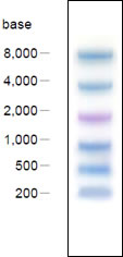
Figure 1. Electrophoresis profile of DynaMarker® Prestain Marker for RNA High (6 μl) on 0.8 % agarose – 2.2 M formaldehyde gel / 1 × MOPS buffer as running buffer.

Figure 2. Electrophoresis profile of DynaMarker® RNA High(1) and DynaMarker® Prestain Marker for RNA High(2) on 0.8 %, 1.0 % and 1.5 % agarose - 2.2 M formaldehyde gel / 1x MOPS buffer as running buffer

Figure 3. Left: Electrophoresis profile of DynaMarker® Prestain Marker for RNA High (1)and RNA sample (2) on 0.8 % agarose - 2.2 M formaldehyde gel / 1x MOPS buffer as running buffer.
Right: Blotting of (1) and (2) onto nylon membrane.
Features
* Prestained ! :
DynaMarker Prestain Marker for Small RNA Plus are highly visible indicators with dual colors of blue and red.
DynaMarker Prestain Marker for RNA High is a highly visible indicator with dual colors of blue and purple.
*From low to high base ! :
The apparent sizes of the bands in the...
DynaMarker Prestain Marker for Small RNA Plus are 20, 30, 40, 50, 75 and 100 bases in length.
The apparent sizes of the bands in the...
DynaMarker Prestain Marker for RNA High are 200, 500, 1,000, 2,000, 4,000 and 8,000 bases in length.
* Ready to use ! :
The DynaMarker Prestain Markers for RNA are ready-to-use mixture. Do not require heat treatment or denaturing agents.
* Visible during electrophoresis
Storage buffer
40 mM MOPS (pH 7.0), 10 mM AcONa, 1 mM EDTA?2Na, 10 % Glycerol
Quality Control
After 24 hrs incubation of the DynaMarker® Prestain Marker for RNA High at 37 ℃, no visible degradation of the marker is observed in 1 % agarose – 2.2 M formaldehyde gel electrophoresis.
Recommended loading volumes
Comb Load volume
4 ~ 6 mm 4 ~ 6 μl
>6 mm >6 μl
Note
- For accurate electrophoretic determination of molecular weights, the DynaMarker® RNA High (code # DM160) or DynaMarker® RNA Easy Measurement N (code # DM170) should be used.
- The migration of the DynaMarker Prestain Marker for RNA High has been optimized to use 0.8-1.5 % of agarose gel concentration (see table 1).
- This product is not for acrylamide gel electrophoresis.
- Particularly avoid freeze – thaw cycle.
|
RNA High |
0.8% |
1.0% |
1.5% |
|
8000 base |
96.5 |
94.8 |
90.4 |
|
4000 |
98.5 |
100.0 |
92.2 |
|
2000 |
104.4 |
102.8 |
101.6 |
|
1000 |
103.4 |
103.2 |
101.4 |
|
500 |
106.4 |
102.0 |
103.1 |
|
200 |
102.7 |
100.0 |
108.7 |
Recommended usage
The DynaMarker® Prestain Marker for RNA High is suitable for monitoring denaturing agarose gel electrophoresis and blotting onto membrane. One example is shown below:
Electrophoresis and blotting of DynaMarker® Prestain Marker for RNA High
1) Preparation of 0.8 % agarose – 2.2 M formaldehyde gel
Agarose 0.8 g
10 × MOPS 10 ml
deionized formaldehyde 18 ml
RNase free water 72 ml
100 ml
Dissolve the agarose by boiling in a microwave oven. Cool the solution to 55 ℃ and add 10 ml of 10 × MOPS buffer and 18 ml of deionized formaldehyde. In a fumehood, cast an agarose gel with slots formed by a 4~6 mm comb. Remove the comb, place the gel in the gel tank, and add sufficient 1 × MOPS running buffer to cover to a depth of ~1 mm.
2) Loading and electrophoresis.
Thaw the DynaMarker® Prestain Marker for RNA High completely before use. Load your denatured RNA sample and 6 μl of DynaMarker® Prestain Marker for RNA High (use a 4~6 mm comb) on to a well and run the gel using 1 × MOPS electrophoresis buffer at 4~5 V / cm.
3) Transfer the DynaMarker® Prestain Marker for RNA High and RNA from the gel to positively charged
nylon membranes. (see figure 3 and 4)
3-1) Place the gel in RNase-free glass dish, and rinse (for formaldehyde removal) with several changes of sufficient deionized water to cover the gel.
3-2) Add ~10 gel volumes of 3 M NaCl / 10 mM NaOH (transfer buffer) to the dish and soak for 30 min.
3-3) Cut a piece of nylon membrane slightly larger than the gel. Soak the membrane and two sheets of
blotting paper of appropriate size in 10× SSC for at least 5 min.
3-4) Place the support (e.g. oblong sponge) in a glass or a plastic dish. Fill the dish with enough transfer buffer (soak the support about half-submerged in buffer.).
3-5) Place the gel on the support in inverted position so that it is centered on the wet blotting paper.
3-6) Place the wet nylon membrane on top of the gel. (! notice: Remove air bubbles.)
3-7) Place the wet blotting paper on the top of the wet nylon membrane. (! notice: Remove air bubbles.)
3-8) Cut a stack of paper towels (5~8 cm high), and place the towels on the blotting papers.
3-9) Put a glass plate on the top of the stack and weight it down with an about 400 g weight.
3-10) Allow upward transfer of RNA to occur for 1hr.
3-11) Transfer the membrane to a glass tray containing 6× SSC, and rinse for 5 min.
3-12) Remove the membrane from the 6× SSC and allow excess fluid to drain away. Then dry the
membrane on blotting paper for a few minutes.
3-13) Fix the RNA to the membrane with a UV-crosslinker.
3-14) Cut off the marker lane.
3-15) Carry out northern hybridization.
Electrophoresis of Dyna in 64 x speed
References
- Joseph Sambrook, and David W. Russell (2001) Molecular Cloning: A Laboratory Manual, 3rd ed., Cold Spring Harbor Laboratory Press.
- Frederick M. Ausubel, Roger Brent, Robert E. Kingston, David D. Moore, J. G. Seidman, John A. Smith, and Kevin Struhl (1994-) Current Protocols in Molecular Biology, John Wiley & Sons, Inc.
- Virus Research Volume 185, 24 June 2014, Pages 92–102
- Hu, S., Kim, H., Yang, P., Yu, Z., Ludeke, B., Mobilia, S., ... & Abraham, J. (2025). Structural and functional analysis of the Nipah virus polymerase complex. Cell, 188(3), 688–703.
Related Products
Please, click below each image if you need more info.
 |
|
 |
 |
 |
 |
 |
|
 |
 |
