

Drebrin antibody (M2F6-Shirao) Mouse mAb
Product#: FNK-AAB002
$908.23
Availability:
Ships in 1-2 Weeks
Drebrin antibody (M2F6-Shirao) Mouse mAb
Catalog No.: FNK-AAB002
Size: 100 uL
Storage: -20℃ (For long-term, make aliquots and store below -20°C)
**Do not repeat freeze and thaw, as the antibody may deteriorate and not perform as intended.
Background Information:
Drebrin is an actin-binding protein that stabilizes actin filaments and has a role in cell migration, adhesion, neurite formation, endocytosis, etc. Moreover, drebrin-decorated stable actin filaments accumulate in dendritic spines and are crucial in synaptic plasticity.
M2F6 antibody is secreted from mouse hybridoma clone M2F6 established by Dr. Shirao in 1986 (ref. 5) and has been used as a worldwide de facto standard in drebrin research. We have improved the quality of the M2F6 antibody and rebranded it as the M2F6-Shirao® antibody. The epitope of this antibody is conserved from birds to humans, and the antibody strongly reacts with non-degraded drebrin.
Antigen: Purified Chicken Drebrin E
Host Species: Mouse
Clone Name: M2F6-Shirao®
Isotype: IgG1
Purification Method: Protein A purified
Storage Buffer: PBS with 1% BSA, 0.05% NaN3
Specificity: Human/Cat/Mouse/Rat/Chicken. React with drebrin E and A (product of drebrin gene DBN1).
Applications: IHC (suitable for paraffin/frozen sections), ICC, WB, IP
Recommended Dilutions:
- IHC 1:100-1:1000
- ICC, WB 1:1000
- IP 1-5 µg
What is Drebrin?
Drebrin was originally reported in chicken brains (Shirao et al., 1984) and named for its developmentally regulated expression in the brain (Developmentally REgulated BRain proteIN). It is an actin-binding protein involved in neuronal cytoskeletal dynamics and is classified into two major isoforms, Drebrin E and Drebrin A, which are generated by alternative splicing of a single DBN1 gene. Both Drebrin A and Drebrin E share a common domain structure at their N-terminus. While the amino acid sequences of Drebrin A and Drebrin E are nearly identical, only Drebrin A contains an additional sequence known as Ins2. Drebrin E is primarily expressed during fetal and juvenile stages, whereas Drebrin A is specifically expressed in neurons at the mature stage.
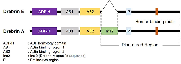
Figure. Structure of human Drebrin
Drebrin E accumulates extensively in axonal growth cone of neurons, affecting neurite outgrowth and axon growth. As neurons mature, Drebrin E disappears and Drebrin A comes to appear instead. Drebrin A accumulates at the heads of dendritic spines and its localization is known to change in response to NMDA receptor activity. This Drebrin dynamics plays a crucial role in synaptic plasticity, the fundamental mechanism underlying learning and memory. In Alzheimer’s disease associated with memory impairment, postmortem analyses of human brain tissue have shown that Drebrin levels are already reduced at the mild cognitive impairment (MCI) stage. In addition, in vitro studies have demonstrated an amyloid-β (Aβ) oligomer–dependent decrease in Drebrin.Features
- This antibody is derived from clone M2F6, established by Shirao et al. who reported Drebrin first.
- Drebrin antibody (clone: M2F6) has been used as the global de facto standard in Drebrin research.
- It is useful as a marker for dendritic spines in mature neurons or as a marker for evaluating neuronal maturation.
Specifications
- Host species: Mouse
- Clone name: M2F6
- Immunogen: Purified Chicken Drebrin E *1
- Reactivity: Human, Mouse, Rat, Chicken, Cat
- Application: IC、IHC(frozen or paraffin sections), IP, WB *2
- Isotype: IgG1
*2 IC: Immunocytochemistry, IHC: Immunohistochemistry, IP: Immunoprecipitation, WB: Western blotting
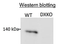
Verification of antibody specificity
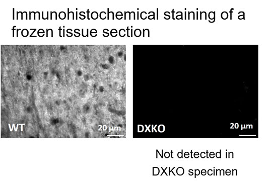
The specificity of this antibody was verified by immunohistochemical staining (lower left panels) and Western blotting (lower right panel) using Drebrin knockout mice.(Specimen: Mouse cerebral cortex, WT: Wild type mouse, DXKO: Drebrin knockout mouse)
 |
 |
Application Examples
Cell staining: Staining of developing primary neurons from rat hippocampus
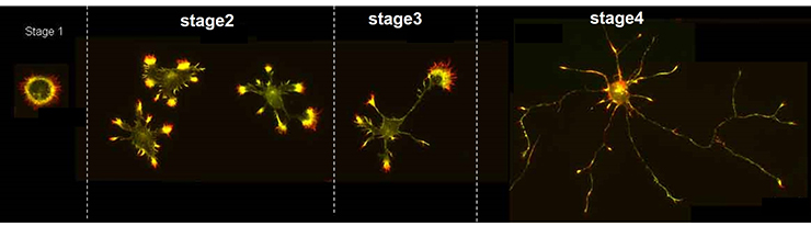
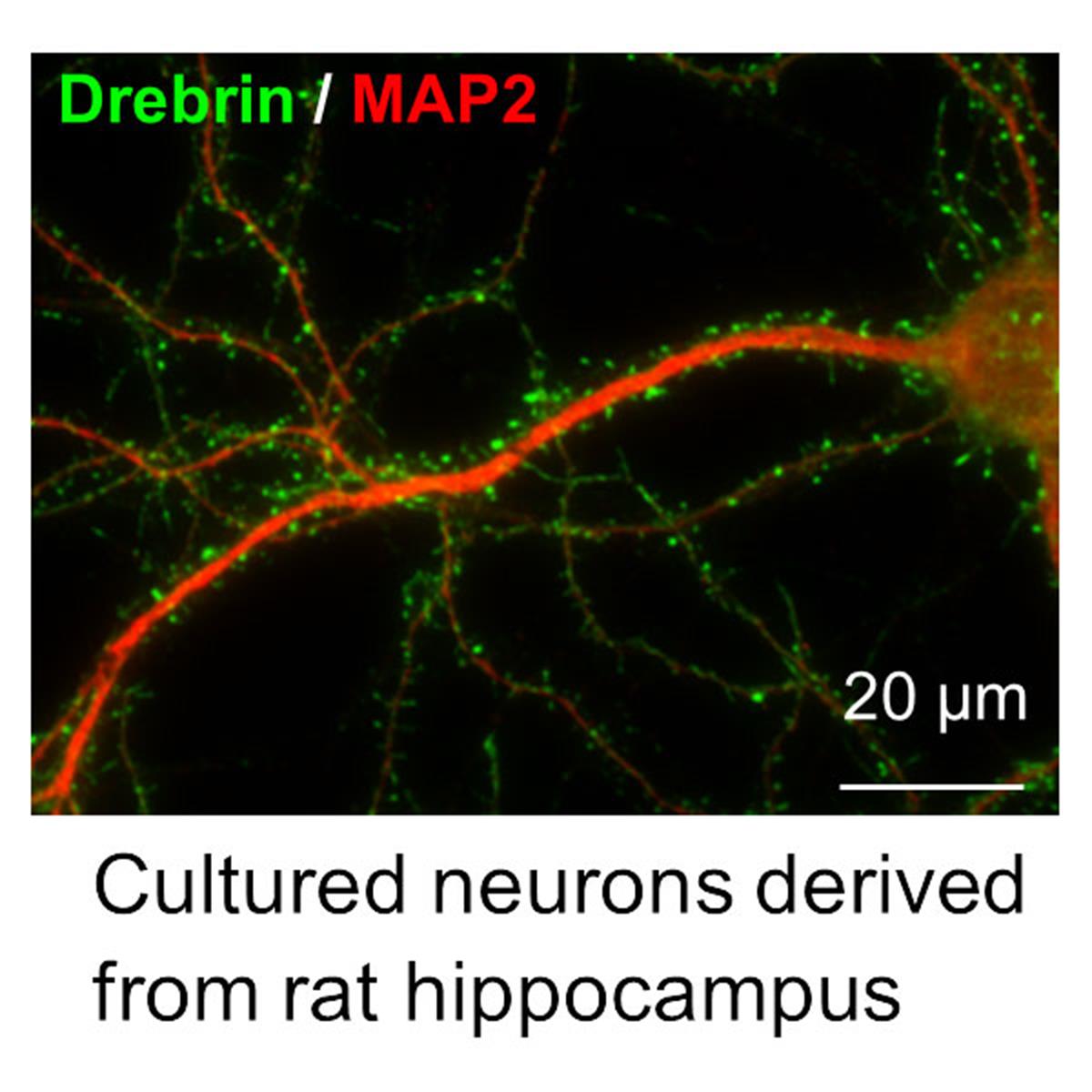
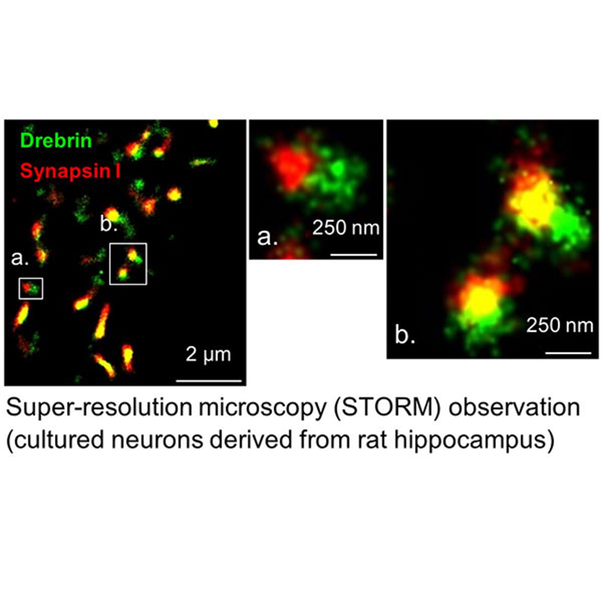
Localization of Drebrin in primary rat hippocampal neurons was observed.
Staining of developing neurons (Stage 1-4) with this antibody visualized Drebrin accumulation at the growth cones of neurite tips (Green: Drebrin, Red: F-actin).
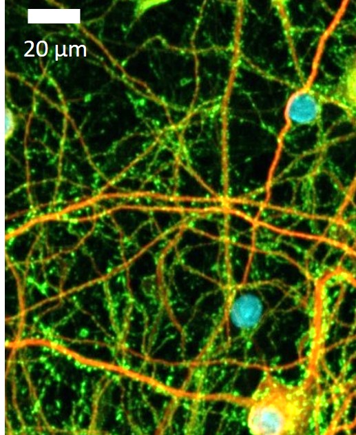
At the maturation stage of neurons (day 21), Drebrin accumulation is observed in dendritic spine heads.
(Green: Drebrin, Red: MAP2, Blue: DAPI)
Cell staining: Staining of human iPS cell-derived neurons
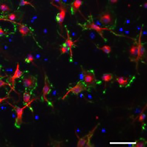
As seen in the case of primary cultured neurons, Drebrin accumulates at the growth cones of neurite tips in immature, early-stage (3 weeks post-culture) human iPS cell-derived neurons, similar to primary cultured neurons.
(Red: MAP2, Green: Drebrin, Blue: DAPI)
For mature human iPS cell-derived neurons, please refer to the following publication.
Reference: Lin, W., et al., iScience, 26(4):106285 (2023). [PMID: 37034988]
Immunohistochemical staining: Paraffin sections of human cerebral cortex
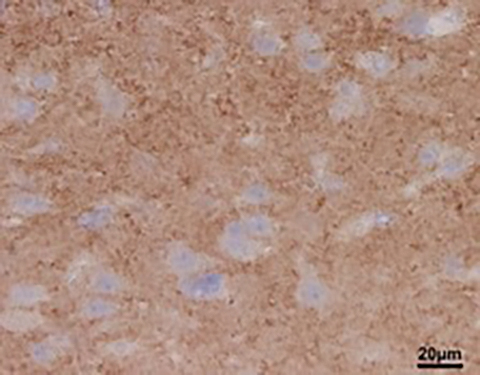
Drebrin accumulation in spines (granular staining) are visible as granular staining. (Provided by Professor Yokoo, Gunma University)
Immunohistochemical staining: Expression of Drebrin in the Alzheimer's disease brain
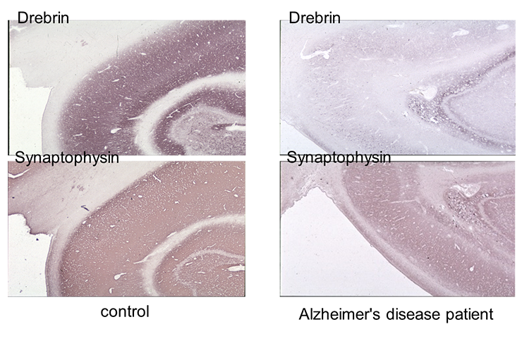
Immunohistochemical staining of Human hippocampal tissue specimens from Alzheimer's disease patients was performed using this antibody.
Although expression of Synaptophysin, a presynaptic marker, was not significantly changed, expression of Drebrin was reduced in patient brains.
Western Blot: Expression of Drebrin in Alzheimer's Disease Brain
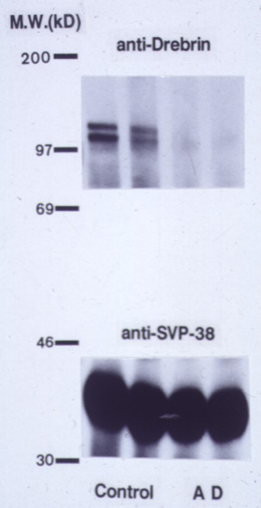
Western blot analysis revealed reduced expression of Drebrin in Alzheimer's disease patients, while synaptophysin, a presynaptic marker, did not show significant expression change.
Top panel: Drebrin, Bottom panel: Synaptophysin
Left two lanes: Control specimens, Right two lanes: Specimens from Alzheimer's disease patients
Publications
- Zhao, M., et al., Neural Regen. Res., 19(7):1602-1607 (2024). [PMID: 38051905]
- Takahashi, K., et al., Sci. Rep., 14(1):11519 (2024). [PMID: 38769131]
- Yamazaki, H., et al., Neurosci. Res., 199:30-35 (2024). [PMID: 37659612]
- Kreis, P., et al., Nat. Commun., 10(1):486 (2019). [PMID: 30700723]
- Counts, S.E, et al., J. Neuropathol. Exp. Neurol., 65(6):592-601 (2006). [PMID: 16783169]
- Shirao, T. and Obata, K., Brain Res., 394(2):233-244 (1986). [PMID: 3768727]
- Shirao, T. and Sekino, Y., (Eds.) (2017) Drebrin. (Advances in Experimental Medicine and Biology, vol 1006) Tokyo, Springer
NOTE: This product is for research use only. Do not use for commercial purposes such as disease diagnosis, treatment or prevention, or sale to third parties.