

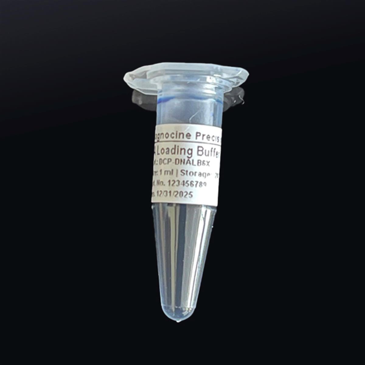
DNA Loading Buffer [6X]
FluxMPS™ DNA Loading Buffer [6X]
An MPS-grade, sucrose-based 6X DNA loading buffer engineered for high-resolution agarose gel electrophoresis. Dual bromophenol blue and xylene cyanol tracking dyes give real-time migration visibility, while quadruple-stage 0.1 µm / 0.04 µm filtration delivers the ultra-clean, DNase- and RNase-free purity molecular biology and microfluidic sample-prep workflows demand.
- Filtered 0.1-micron membrane twice and 0.04-micron membrane twice for ultra-clean, mycoplasma-scale purity
- Sucrose-based density loading buffer at pH 7.4 for sharp, well-resolved DNA bands
- Contains bromophenol blue and xylene cyanol dual tracking dyes for migration monitoring
- Manufactured with Ultrapure Type 1 water (18.2 MΩ·cm) under ISO Class 5 (Class 100) fill
- No DNase or RNase activity detected after 18-hour incubation at room temperature
- ISO 13485-certified, CE-approved manufacturing with final QA at DiagnoCine's Totowa, New Jersey facility
- Customizable concentrations, tracking dyes, and pH available on request
- pH7.4
- Sucrose40%
- Bromophenol Blue0.25%
- Xylene Cyanol0.25%
- Tris-HCl10 mM
- EDTA60 mM
- Filtration0.1 µm x2 + 0.04 µm x2
- DNase ActivityNone Detected
- Storage-20°C
- Shelf Life1 Year
Engineered where standard loading buffers fail
Conventional gel loading dyes are typically filtered once through a 0.22 µm membrane, leaving subvisible particulate and inconsistent tracking-dye purity that can distort band resolution or interfere with downstream imaging. FluxMPS™ DNA Loading Buffer [6X] is built around a quadruple-stage filtration architecture and validated DNase/RNase-free performance.
Microchannel-safe purity
Final 0.04 µm filtration stage removes fine particulate that a single 0.22 µm pass would allow through, supporting clean loading into gel wells and fine-bore microfluidic dispensers.
Precise, stable pH
Formulated at pH 7.4 with a Tris-HCl and EDTA base to keep DNA samples stable and density-shifted for clean well loading.
Ultrapure-grade water
Manufactured with Ultrapure Type 1 water (18.2 MΩ·cm), aligned with USP <85> water-quality expectations for sensitive molecular biology reagents.
Clean visualization performance
Validated free of detectable DNase and RNase activity so bromophenol blue and xylene cyanol tracking dyes deliver consistent migration visualization without nuclease interference.
Defined, traceable composition
Every component — sucrose, Tris-HCl, EDTA, and both tracking dyes — is manufactured to a defined concentration and traceable to DiagnoCine's quality system.
Customization on demand
Alternate concentrations, additional tracking dyes, different pH, and other modifications can be requested at support@diagnocine.com.
Quadruple-stage filtration system
DCP-DNALB6X is filtered 0.1-micron membrane twice and 0.04-micron membrane twice in a sterile environment, delivering purity well beyond a conventional single 0.22 µm pass and preventing mycoplasma-scale contamination in the finished buffer.
-
1
0.1 µm Pre-filtration I
Removes larger particulate and aggregates from the sucrose, dye, and buffer salt solution ahead of downstream polishing.
-
2
0.04 µm Pre-filtration II
Retains finer particulates and bioburden that a 0.1 µm pass alone would pass through.
-
3
0.1 µm Sterile-filtration I
A second 0.1 µm pass provides redundant clarification ahead of the final polishing stage.
-
4
0.04 µm Sterile-filtration II — Final Polish
A second 0.04 µm pass performed in a sterile environment gives the finished buffer its final polish prior to aseptic fill.
Performance vs. conventional loading buffer
Sequential 0.1 µm and 0.04 µm filtration, applied twice each, removes finer particulate than a single conventional 0.22 µm pass can achieve, supporting clean gel loading and downstream imaging.

© Diagnocine® — DCP-DNALB6X
Where DCP-DNALB6X is used
Formulated for loading DNA samples into agarose gel wells, DCP-DNALB6X also supports the broader molecular biology sample-prep steps behind microfluidic, organ-on-a-chip, and cell-model research.
Automated Bioreactors & Robotics
An optional 0.01 micron (10 nm) ultra-filtered variant of this DNA Loading Buffer [6X] is available for automated liquid-handling and robotic gel-loading platforms, protecting fine-bore dispensing needles and optical sensors from subvisible particulate beyond what standard 0.1/0.04 micron filtration removes.
- Total Particulate Exclusion: further reduces subvisible particulate for automated dispensing lines.
- Valve & Sensor Protection: helps protect microvalves and optical detection components from fouling.
- Extended Perfusion Stability: supports consistent long-run performance in automated liquid-handling systems.
Inquiry Required: contact support@diagnocine.com to request the 0.01 µm ultra-filtered grade.
Micro Physiological System (MPS) & Chip
Confirms plasmid and construct identity by gel electrophoresis before cell lines are engineered for organ-on-a-chip and tissue-chip platforms.
Loading, Migration & Band Resolution
Increases sample density with sucrose to sink DNA samples into gel wells cleanly, preventing diffusion and loss during loading.
iPSC-Derived Model Handling
Supports genotyping and clone verification of iPSC-derived lines by gel electrophoresis prior to downstream differentiation studies.
Endothelial & Primary Cell Perfusion
Verifies vector and plasmid identity used to transduce endothelial and primary cell models ahead of perfusion-based experiments.
ELISA, Blotting & Blocking
Confirms probe and construct DNA integrity by gel electrophoresis before use in blotting-based detection workflows.
Microscopy & Optical Sensing
Verifies reporter construct integrity by gel electrophoresis prior to imaging experiments on confocal and biosensor-based chip platforms.
Full specification sheet
Every value below is quality-controlled and lot-verified for DCP-DNALB6X.
| Parameter | Specification |
|---|---|
| Formulation | Sucrose-based density loading buffer with bromophenol blue and xylene cyanol tracking dyes |
| Appearance | Blue-purple or violet color |
| pH (USP <791>) | 7.4 |
| Sucrose concentration | 40% |
| Bromophenol blue concentration | 0.25% |
| Xylene cyanol concentration | 0.25% |
| Tris-HCl concentration | 10 mM |
| EDTA concentration | 60 mM |
| Parameter | Specification |
|---|---|
| Sterility USP <71> | Filtered 0.1-micron membrane twice and 0.04-micron membrane twice in a sterile environment |
| DNase activity | None detected after incubation of plasmid DNA for 18 hours at room temperature |
| RNase activity | None detected after incubation of ribosomal RNA for 18 hours at room temperature |
| Water quality | Ultrapure Type 1 water (18.2 MΩ·cm) |
| Manufacturing standard ISO 13485:2016 | ISO 13485-certified, CE-approved facility |
| Fill environment | ISO Class 5 (Class 100) cleanroom |
| Parameter | Specification |
|---|---|
| Storage temperature | -20°C |
| Shelf life | 1 year |
| Parameter | Specification |
|---|---|
| Manufacturing QMS | ISO 13485-certified |
| Regulatory alignment | CE-approved manufacturing facility |
| Production method | Final packaging, quality assurance, and testing performed at the DiagnoCine R&D and Quality Testing Center; customization assembly performed at DiagnoCine Precision, Totowa, New Jersey, USA |
| Intended use | For Research Use Only (RUO). Not intended for clinical, diagnostic, or therapeutic use in humans. |
Full composition
Every component below is quality-controlled to the concentration shown for each lot of DCP-DNALB6X.
| Component | CAS Number | Concentration |
|---|---|---|
| Sucrose | 57-50-1 | 40% |
| Bromophenol blue | 115-39-9 | 0.25% |
| Xylene cyanol | 2650-17-1 | 0.25% |
| Tris-HCl | 1185-53-1 | 10 mM |
| EDTA | 60-00-4 | 60 mM |
Manufacturing & compliance
DCP-DNALB6X is manufactured, filled, and tested under a controlled quality system.
ISO 13485:2016 QMS
Manufactured under ISO 13485-certified and CE-approved facilities (suppliers of DiagnoCine Precision).
Ultrapure Type 1 Water
Formulated with Ultrapure Type 1 water (18.2 MΩ·cm) as the base for every batch.
ISO Class 5 Fill & Finish
Final packaging, quality assurance, and testing are completed at the DiagnoCine R&D and Quality Testing Center.
Micro-Batch Precision
Custom formulation requests and assembly are completed at DiagnoCine Precision in Totowa, New Jersey, USA.
DNase / RNase Validation
No DNase activity detected after incubation with plasmid DNA for 18 hours at room temperature; no RNase activity detected after incubation with ribosomal RNA for 18 hours at room temperature.
Sterile Filtration USP <71>
Filtered 0.1-micron membrane twice and 0.04-micron membrane twice in a sterile environment.
Appearance Verification
Each lot is confirmed for the expected blue-purple or violet appearance prior to release.
Documentation / CoA
Certificates of Analysis are available on request for each lot.
How DCP-DNALB6X compares
A side-by-side look at how DCP-DNALB6X's filtration and QC architecture compares to conventional gel loading buffers.
| Parameter | DCP-DNALB6X (FluxMPS™) | Conventional 0.22 µm-Filtered Buffer | Standard Alternative |
|---|---|---|---|
| Dual bromophenol blue + xylene cyanol tracking dyes | check_circle | cancel | cancel |
| Final filtration pore size | 0.04 µm | 0.22 µm | 0.22 µm |
| Number of filtration stages | 4 | 1 | 1 |
| DNase / RNase validated | check_circle | cancel | cancel |
| ISO 13485-certified manufacturing | check_circle | cancel | cancel |
| Ultrapure Type 1 water base | check_circle | cancel | cancel |
| Microfluidic / chip-compatible sample prep | check_circle | cancel | cancel |
| Custom formulation available | check_circle | cancel | cancel |
Frequently asked questions
Answers to common questions about DCP-DNALB6X.
Supporting literature
Curated references relevant to DNA gel loading buffers, sucrose-based density loading, tracking dyes, and filtration-based sterility assurance.
- Lee, P.Y., et al. Agarose gel electrophoresis for the separation of DNA fragments. J. Vis. Exp. 2012. doi:10.3791/3923
- Sambrook, J., Russell, D.W. Molecular Cloning: A Laboratory Manual. Cold Spring Harbor Laboratory Press, 2001. doi:10.1101/pdb.top133
- Voytas, D. Agarose gel electrophoresis. Curr. Protoc. Mol. Biol. 2000. doi:10.1002/0471142727.mb0205as51
- Bustin, S.A., et al. Guidelines for nucleic acid handling and quality control in molecular assays. Clin. Chem. 2009. doi:10.1373/clinchem.2008.112797
- Low, L.A., et al. Organs-on-chips: into the next decade. Nat. Rev. Drug Discov. 2021. doi:10.1038/s41573-020-0079-3
- Ingber, D.E. Human organs-on-chips for disease modelling, drug development and personalized medicine. Nat. Rev. Genet. 2022. doi:10.1038/s41576-022-00466-9
- Uphoff, C.C., Drexler, H.G. Detecting mycoplasma contamination in cell cultures. Methods Mol. Biol. 2011. doi:10.1007/978-1-61779-108-6_1
- Van Meerloo, J., et al. Gel electrophoresis: a review of nucleic acid separation methods. Methods Mol. Biol. 2011. doi:10.1007/978-1-61779-080-5_20