

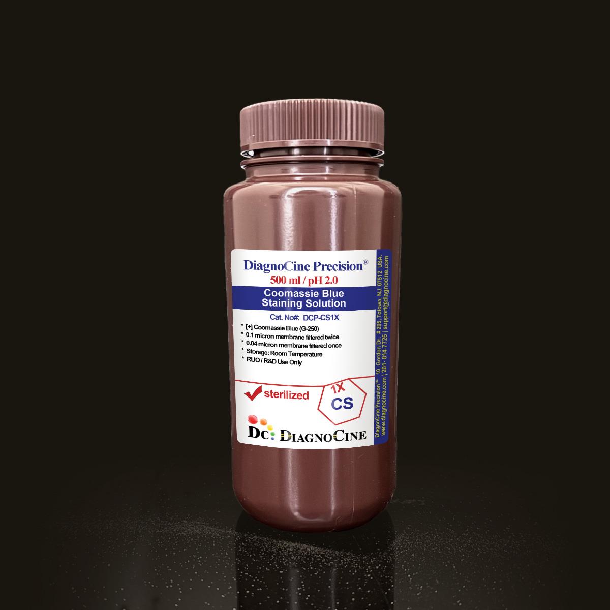
Coomassie Blue Staining Solution
FluxMPS™ Coomassie Blue Staining Solution
An MPS-grade, ultra-filtered colloidal Coomassie G-250 staining solution formulated at pH 2.0 for rapid, high-sensitivity protein visualization in SDS-PAGE gels. Manufactured with quadruple-stage filtration and Ultrapure Type 1 water, DCP-CS1X delivers a sterile, low-particulate reagent suited to microfluidic-adjacent proteomics and mass spectrometry quality-control workflows.
- Quadruple-stage filtration: 0.1 µm membrane twice and 0.04 µm membrane twice for ultra-low particulate purity
- Sterile-filtered in a controlled environment; no DNase or RNase activity detected after 18 hr incubation
- Precise pH 2.0 formulation for consistent colloidal Coomassie G-250 binding
- Prepared with Ultrapure Type 1 water (18.2 MΩ·cm)
- Mass spectrometry-compatible; non-covalent binding allows protein recovery after staining
- Eco-friendly formulation with reduced hazardous waste versus traditional Coomassie R-250 stains
- Supplied as 2 x 500 mL bottles under ISO 13485-certified, CE-approved manufacturing
- Custom concentrations, pH, and additive formulations available on request
- Format2 x 500 mL
- Active DyeCoomassie Blue G-250 (0.1%)
- AppearanceBlue liquid (amber bottle)
- pH2.0
- Filtration0.1 µm x2 + 0.04 µm x2
- DNase / RNaseNot detected
- StorageRoom temperature
- Shelf Life12 months
- Intended UseResearch Use Only (RUO)
- ManufacturingISO 13485 / CE, Totowa, NJ, USA
Engineered where standard Coomassie stains fall short
Conventional 0.22 µm-filtered staining solutions and traditional methanol/acetic-acid R-250 formulations carry subvisible particulates, inconsistent pH, and hazardous waste burdens. DCP-CS1X is built for laboratories that need reproducible, microchannel-safe staining chemistry from the first bottle to the last.
Microchannel-safe purity
Quadruple-stage 0.1 µm / 0.04 µm filtration keeps particulates out of downstream microfluidic and imaging workflows.
Precise, stable pH
Formulated at pH 2.0 for consistent colloidal Coomassie G-250 dye binding, lot after lot.
Ultrapure-grade water
Prepared with Ultrapure Type 1 water (18.2 MΩ·cm) as the aqueous base.
Low background, high reproducibility
Endpoint colloidal staining, non-covalent binding, and a wide linear range support both qualitative and quantitative protein analysis with protein recovery after staining.
Defined, traceable composition
A single active ingredient, Coomassie Blue G-250 at 0.1%, keeps the formulation simple, mass-spec compatible, and easy to QC.
Customization on demand
Alternate concentrations, pH, and additive chemistries are available — contact support@diagnocine.com.
Quadruple-stage filtration system
DCP-CS1X is filtered through a 0.1 µm membrane twice and a 0.04 µm membrane twice, then sterile-filled in a controlled environment — a purity architecture designed to keep the staining solution free of particulates that could interfere with downstream gel imaging, mass spectrometry, or microfluidic sample handling.
-
1
0.1 µm Pre-filtration I
First-pass removal of large particulates and aggregates, extending the life of downstream filters.
-
2
0.04 µm Pre-filtration II
Retention of fine particulates and bioburden ahead of the sterile-filtration stages.
-
3
0.1 µm Sterile-filtration I
Second-pass 0.1 µm filtration provides redundancy ahead of final polishing.
-
4
0.04 µm Sterile-filtration II — Final Polish
Final 0.04 µm polish performed under ISO Class 5 aseptic fill conditions. This quadruple-stage approach is designed to prevent mycoplasma contamination; the smallest mycoplasma type is approximately 0.2 microns.
Performance vs. conventional staining solution
Sequential 0.1 µm and 0.04 µm filtration, applied twice each, removes finer particulates than a single conventional 0.22 µm pass, supporting cleaner downstream SDS-PAGE and mass spectrometry workflows.

© Diagnocine® — DCP-CS1X
Where DCP-CS1X fits your workflow
A high-sensitivity, colloidal protein stain optimized for rapid visualization of proteins in SDS-PAGE gels, designed for researchers prioritizing speed, environmental safety, and downstream compatibility.
Automated Bioreactors & Robotics
For high-throughput and automated gel-staining platforms, an optional 0.01 µm (10 nm) ultra-filtered variant of DCP-CS1X is available to further reduce particulate load on automated fluidics and imaging robotics.
- Total Particulate Exclusion for automated staining instrument fluidics
- Valve & Sensor Protection in robotic gel-handling systems
- Extended Perfusion Stability for continuous-flow staining/destaining protocols
Inquiry Required: the 0.01 µm (10 nm) ultra-filtered grade is available on request — contact support@diagnocine.com.
Micro Physiological System (MPS) & Chip
Post-lysis protein QC for organ-on-a-chip effluent and lysate samples ahead of downstream microfluidic proteomic workflows.
Wash, Dilution & Reconstitution
Compatible with the post-electrophoresis ultrapure-water rinse steps and microwave-assisted staining protocol.
iPSC-Derived Model Handling
Total-protein detection for iPSC-derived lysates prior to gel-based expression profiling.
Endothelial & Primary Cell Perfusion
Total-protein normalization for HUVEC, HAEC, and primary cell lysates run on SDS-PAGE.
ELISA, Blotting & Blocking
Widely used for Western blot total-protein loading normalization alongside ELISA and IHC/IF sample QC.
Microscopy & Optical Sensing
Supports post-assay protein-content verification for samples destined for confocal and biosensor workflows.
Recommended staining protocol
A microwave-assisted protocol enables visible bands within 5–10 minutes.
| Action | Details |
|---|---|
| Post-electrophoresis rinse | Wash gel 3× with ultrapure water (100 mL per wash), 5 min per wash; removes residual SDS and buffers |
| Microwave pre-treatment | Submerge gel in 100 mL water; microwave at 800W for 1 min/gel (1–2 min total); ensures even heating, avoid boiling |
| Staining | Replace water with 50–100 mL staining solution; microwave at 800W for 30 sec/gel; solution turns dark blue, proteins bind dye rapidly |
| Agitation | Shake gently on orbital shaker for 15–30 min; bands should be visible within 5–10 min |
| Destaining | Replace stain with water; microwave at 800W for 1 min/gel, then shake for 1–2 hours; add Kimwipes® to absorb excess dye |
Physical, chemical, and quality parameters
All values below reflect DCP-CS1X as manufactured and tested by Diagnocine.
| Parameter | Specification |
|---|---|
| Formulation / Composition | Coomassie Blue (G-250), 0.1% |
| Appearance | Blue liquid (amber bottle) |
| pH USP <791> | 2.0 |
| Format / Size | 2 x 500 mL |
| Parameter | Specification |
|---|---|
| Sterility | Filtered 0.1 µm membrane twice and 0.04 µm membrane twice in a sterile environment |
| DNase Activity | None detected (plasmid DNA, 18 hr, room temperature) |
| RNase Activity | None detected (ribosomal RNA, 18 hr, room temperature) |
| Water Quality | Ultrapure Type 1 water (18.2 MΩ·cm) |
| Filtration System | 0.1 µm x2 + 0.04 µm x2 (quadruple-stage) |
| Manufacturing Standard ISO 13485 | ISO 13485-certified, CE-approved facilities |
| Fill Environment | Sterile environment |
| Parameter | Specification |
|---|---|
| Storage Temperature | Room temperature |
| Shelf Life | 12 months |
| Handling | Wear gloves (acidic pH) |
| Disposal | Eco-friendly — pour neutralized solution down drain |
| Parameter | Specification |
|---|---|
| Raw Material Sourcing | Sourced from DiagnoCine Precision suppliers |
| Manufacturing QMS | ISO 13485-certified |
| Regulatory Alignment | CE-approved facilities |
| Final Packaging / QA / Customization Location | DiagnoCine Precision, Totowa, New Jersey, USA |
| Intended Use | Research Use Only (RUO) |
Full composition
DCP-CS1X contains a single active dye at defined concentration, manufactured under lot-controlled release.
| Component | CAS Number | Concentration |
|---|---|---|
| Coomassie Blue (G-250) | 6104-58-1 | 0.1% |
Manufacturing & compliance
DCP-CS1X is manufactured, packaged, and tested under a controlled quality system.
ISO 13485:2016 QMS
Manufactured under ISO 13485-certified and CE-approved facilities.
Ultrapure Type 1 Water
Formulated with Ultrapure Type 1 water (18.2 MΩ·cm) as the aqueous base.
ISO Class 5 Fill & Finish
Final 0.04 µm polish and fill performed under ISO Class 5 (Class 100) aseptic conditions.
Micro-Batch Precision
Final packaging, quality assurance, testing, and customization completed at the DiagnoCine R&D and Quality Testing Center in Totowa, New Jersey, USA.
Nuclease Testing (DNase / RNase)
No DNase or RNase activity detected after 18 hr incubation with plasmid DNA and ribosomal RNA at room temperature.
Sterility & Bioburden Control
Quadruple-stage 0.1/0.04 µm filtration performed in a sterile environment to prevent mycoplasma and microbial contamination.
pH Verification (USP <791>)
Each lot is confirmed to formulate at pH 2.0.
Documentation / CoA
Certificate of Analysis available on request — contact support@diagnocine.com.
How DCP-CS1X compares
DCP-CS1X (Coomassie G-250) is compared below against traditional Coomassie R-250 and a generic conventional 0.22 µm-filtered staining solution.
| Parameter | DCP-CS1X (FluxMPS™) | Coomassie R-250 (Standard) | Conventional 0.22 µm-Filtered Stain |
|---|---|---|---|
| Sensitivity | 8–10 ng/band | 50–100 ng/band | Not specified |
| Toxicity | Non-hazardous | Hazardous (MeOH/HOAc) | Not specified |
| Processing Time | 1.5–2.5 hrs | 4–6 hrs | Not specified |
| Filtration Architecture | 0.1 µm x2 + 0.04 µm x2 | Not specified | Single 0.22 µm pass |
| Mass Spectrometry Compatible | check_circle | cancel | cancel |
| Non-Covalent (Protein Recoverable) | check_circle | cancel | cancel |
| Eco-Friendly Disposal | check_circle | cancel | Not specified |
| Manufacturing QMS | ISO 13485 / CE | Not specified | Not specified |
| Custom Formulation Available | check_circle | cancel | cancel |
Frequently asked questions
Common questions about DCP-CS1X specifications, handling, and customization.
Supporting literature
Curated references on Coomassie staining chemistry, SDS-PAGE workflows, and microfluidic sample handling.
- Neuhoff, V. et al. Improved staining of proteins in polyacrylamide gels including isoelectric focusing gels with clear background at nanogram sensitivity using Coomassie Brilliant Blue G-250 and R-250. Electrophoresis. doi:10.1002/elps.1150090913
- Kang, D. et al. A colloidal Coomassie Blue staining method for proteins separated by polyacrylamide gel electrophoresis. Proteomics. doi:10.1002/1615-9861(200210)2:10<1441::AID-PROT1441>3.0.CO;2-4
- Candiano, G. et al. Blue silver: a very sensitive colloidal Coomassie G-250 staining for proteome analysis. Electrophoresis. doi:10.1002/elps.200305844
- Bull, J.M. et al. Considerations for total protein staining as a loading control alternative to housekeeping proteins in Western blotting. J Vis Exp. doi:10.3791/55519
- Sackmann, E.K. et al. The present and future role of microfluidics in biomedical research. Nature. doi:10.1038/nature13118
- Bhatia, S.N. & Ingber, D.E. Microfluidic organs-on-chips. Nat Biotechnol. doi:10.1038/nbt.2989
- Righetti, P.G. & Chiari, M. Coomassie staining protocols for high-resolution protein gel electrophoresis. Methods Mol Biol. doi:10.1385/1-59259-651-4:1
- Aebersold, R. & Mann, M. Mass-spectrometry-based proteomics. Nature. doi:10.1038/nature01511
- Low, T.Y. et al. Recognizing the potential of H2020 sample preparation standards for proteomics. J Proteome Res. doi:10.1021/acs.jproteome.9b00429