Cell Proliferation
Select CellProliferation
- see the table below
Storage and Handling Conditions
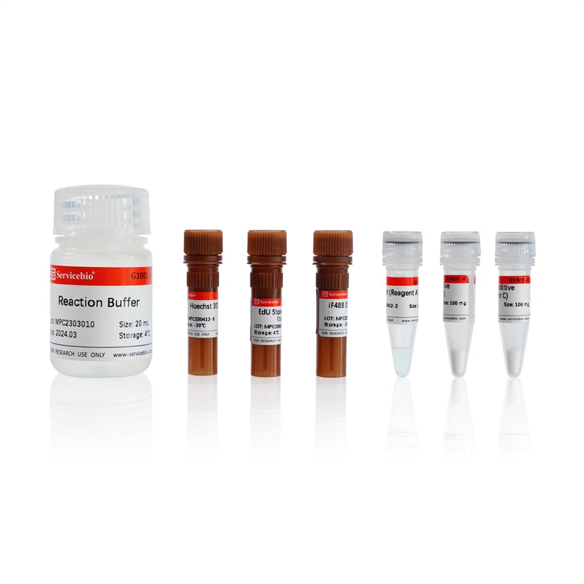
Reagent B(iF488 dye) should be store at -20°C away from light;
Reagent C should be store at -20°C,subpackage after dissolved;
EdU storage solution can be store at -20°C;
EdU catalytic reagent (Reagent A) and reaction buffer can be stored at 4°C;
Valid for one year.
| Cat.No: | Description | Size | Volume | Link |
| SB-G4104-500T | MTT | 500T | 5×1 mL |  |
| SB-G1602 | Click-iT EdU-555 | 100T | 100 rxn | |
| SB-G1603 | Click-iT EdU-594 | |
||
| SB-G1604 | Click-iT EdU-647 | |
||
| SB-G1601 | Click-iT EdU-488 | |
Description/Introduction:
Analyzing cell proliferation ability is a common and important evaluation method in life sciences. It can judge the influence of certain genes, drugs, etc. on cells cultured in vitro, or analyze the growth and renewal ability of tissue cells under different conditions or stimulation. At present, there are many methods to detect cell proliferation. Most of them use some metabolic enzymes produced by cells to indirectly assess cell proliferation activity (such as CCK-8 method, MTT method, etc.), but some drugs or the state of the cell itself will have a certain impact on the results of the assessment. Direct detection of DNA synthesis in cells to determine cell proliferation is recognized as the most accurate and effective detection method. However, both the original radiolabeled nucleoside incorporation method and the subsequent improvement of the BrdU method based on antibody detection have their own limitations.
EdU (5-Ethynyl-2'-deoxyuridine, 5-ethynyl-2'-deoxyuridine) is a thymidine analogue containing an acetylene group, when injected into animals or incubating cells cultured in vitro, these small molecules can quickly diffuse to various organs and tissues, and infiltrate into the cells, and can replace thymidine (T) into newly synthesized DNA during cell proliferation. The acetylene group in the EdU molecule can react with the fluorescently iF488 labeled azide compound probe to form a stable triazole ring under the catalysis of copper ions, so the newly synthesized DNA can be labeled with the corresponding fluorescent probe. Compared with the radiolabeled nucleoside incorporation method, the EdU detection method has no limiting factors such as radioactive contamination; compared with the BrdU detection method, the EdU detection method does not require DNA denaturation or antigen-antibody reaction, which greatly reduces the complexity of the experiment and also make the experiment more time-saving, more sensitive, more stable and more accurate.
This kit can be used to detect cell proliferation in cultured cells or animal tissues. The fluorescent probe in this kit is green fluorescence, the maximum excitation wavelength is 491 nm, and the maximum emission wavelength is 516 nm. After the proliferating cells are labeled, the cell nucleus will show bright green fluorescence, and the cell nucleus will be jointly labeled with the matching conventional nuclear dye (This kit provides Hoechst 33342 cell nuclear dye), you can use fluorescence microscope, laser confocal microscope and other instruments to directly observe cell proliferation; you can also use flow cytometry to detect the fluorescence intensity of cultured cells in vitro, and then determine the cell cycle based on the fluorescence intensity DNA replication activity in mid-S phase.