Can Get Signal ® immunostain
Can Get Signal ® immunostain
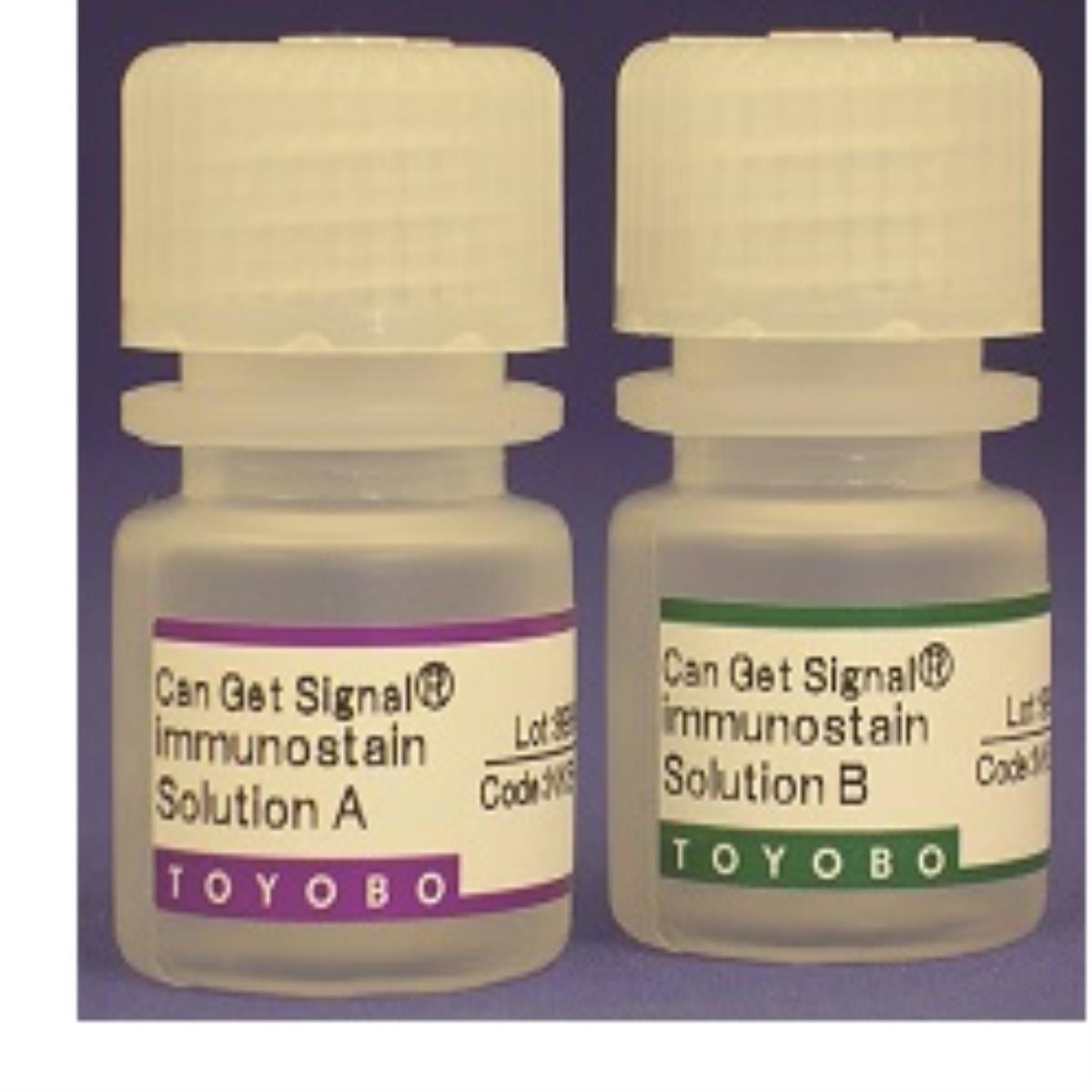
Can Get Signal® immunostain is a reaction solution that contains an accelerator for antigen-antibody reactions, which improves sensitivity, specificity, and S/N of immunohistochemistry (IHC) and immunocytochemistry.
| Product Name | Cat. No. | Size | Storage | Order |
|---|---|---|---|---|
| Can Get Signal ® immunostain | TYB-NKB-401 | Solution A(5 ml)+ Solution B(5 ml) | 4°C | Current page |
DESCRIPTION
Can Get Signal® immunostain is a reaction solution that contains an accelerator for antigen-antibody reactions, which improves sensitivity, specificity, and S/N of immunohistochemistry (IHC) and immunocytochemistry.
FEATURES
- Improves sensitivity, specificity, and S/N of IHC.
- Can be applied to various detection systems (e.g., chromogenic, chemiluminescence, or fluorescence).
- Can be used with ABC or polymer complex methods.
- Solutions A and B exhibit various properties for improving results.
- Can be used directly without dilution (Ready-to-use).

APPLICATIONS
- Immunohistochemistry (IHC)
- Immunocytochemistry
COMPONENTS
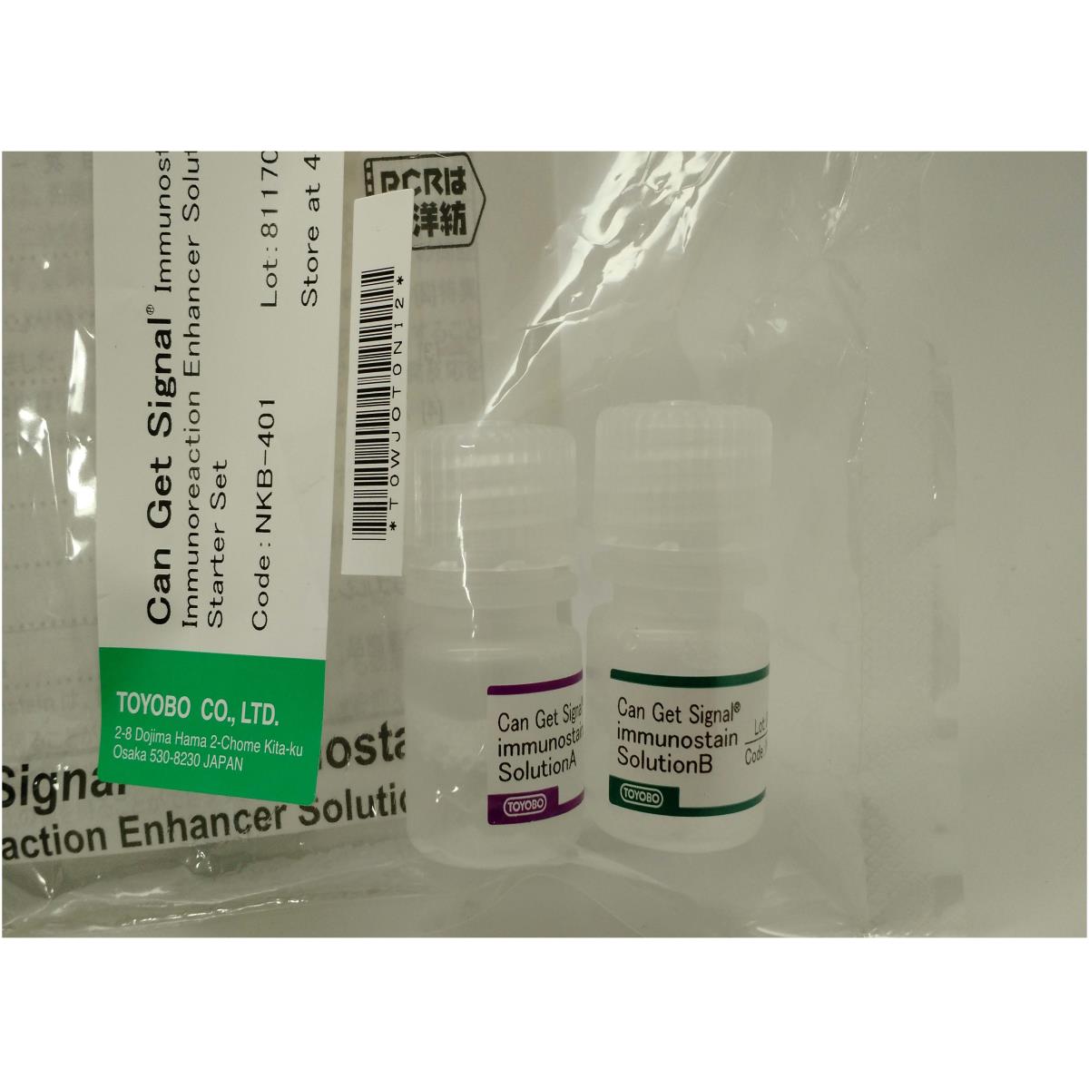
This kit includes the following components. All reagents should be stored at 4ºC and protected from light.
| Reagent Name | Code No. | ||
|---|---|---|---|
| TYB-NKB-401 | TYB-NKB-501 | TYB-NKB-601 | |
| Solution A | 5 ml | 20 ml | - |
| Solution B | 5 ml | - | 20 ml |
| Link | current page |  |
|
TYPICAL REACTION FLOW
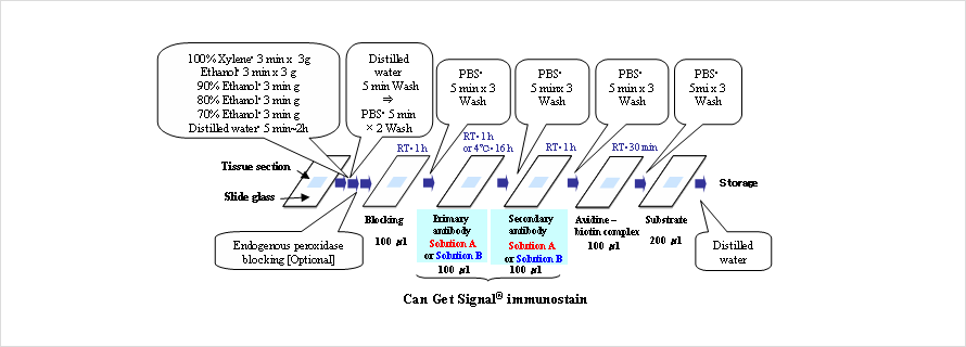
Flow chart of immunostaining with Can Get Signal® immunostain
APPLICATION DATA
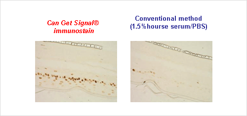
Example 1. Detection of PCNA using paraffin-embedded sections
The localization of PCNA (proliferating cell nuclear antigen) expression in human skin was detected using paraffin-embedded sections of the human skin tissue model TESTSKINTM (Toyobo). Detection was performed by the ABC method with anti-PCNA mouse monoclonal antibody as the primary antibody and biotinylated mouse IgG as the secondary antibody. Each antibody was diluted with Solution A of Can Get Signal® immunostain prior to use. As a control experiment, PBS(-) containing 1.5% normal horse serum (conventional method) was used instead of Solution A of Can Get Signal® immunostain. As a result, Can Get Signal® immunostain produced higher signals and lower background than the conventional method.
Example 2. Detection of paxillin with the fluorescent antibody method
The localization of paxillin in Swiss 3T3 cells was detected using anti-paxillin polyclonal antibody and Alexa488-conjugated rabbit IgG antibody. As a result, the exposure time for detection could be reduced from 3 s to 1 s by using Can Get Signal immunostain®, and the detailed fiber structure (fibrillar adhesion) could also be detected by using Can Get Signal® immunostain. However, the structure could not be detected with the conventional method due to high background signals.
Fig. 2C shows a merged picture of the immunological detection with anti-paxillin and anti-phospho-tyrosine antibodies, and actin staining.
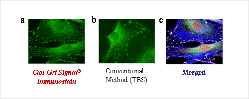
Fig. 2. Immunocytochemistry detection of paxillin with the fluorescent antibody method
*The data was kindly provided by Dr. Harada, Tokyo Institute of Technology.