

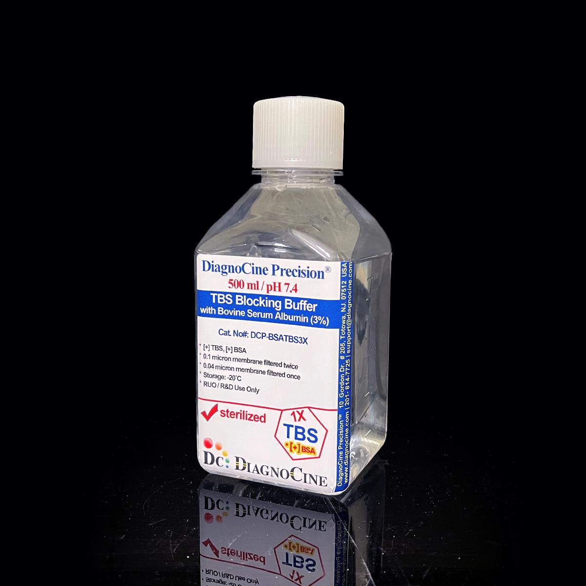
TBS Blocking Buffer with Bovine Serum Albumin (3%)
FluxMPS™ TBS Blocking Buffer with Bovine Serum Albumin (3%)
An MPS-grade Tris-Buffered Saline blocking reagent formulated with 3% Bovine Serum Albumin (BSA) for precise, stable pH and defined molarity. Manufactured through a quadruple-stage 0.1 µm and 0.04 µm filtration architecture using Ultrapure Type 1 water, this phosphate-free buffer minimizes non-specific binding and background interference in Western blot, ELISA, and immunohistochemistry workflows, and is microchannel-safe for organ-on-a-chip (OoC) and microfluidic applications.
- Quadruple-stage purity: filtered through a 0.1 µm membrane twice and a 0.04 µm membrane twice for sterile, ultra-clean blocking buffer.
- Formulated with 3% Bovine Serum Albumin (BSA) in Tris-Buffered Saline (TBS) for high-affinity, low-cross-reactivity blocking.
- Phosphate-free chemistry — compatible with alkaline phosphatase-based detection systems.
- Manufactured with Ultrapure Type 1 water (18.2 MΩ·cm).
- DNase and RNase activity: none detected.
- Stable pH 7.4 formulation supports protein and antibody integrity throughout assay incubation.
- Custom concentrations, pH, and additive formulations available on request.
- pH7.4
- Buffer SystemTris-Buffered Saline (TBS)
- Molarity (Tris-Base)50 mM
- Ionic Strength (NaCl)150 mM
- Blocking Agent (BSA)3%
- DNase / RNase ActivityNone detected
- Sterility0.1 µm x2, 0.04 µm x2
- AppearanceClear, colorless liquid
- Storage-20°C
- Shelf Life2 years
Engineered where standard blocking buffers fail
Conventional 0.22 µm-filtered blocking buffers can carry subvisible particulates, inconsistent BSA quality, and pH drift that elevate background signal in sensitive immunoassays and accumulate in microfluidic channels. FluxMPS™ TBS Blocking Buffer with BSA (3%) is engineered to remove these failure modes at the source.
Microchannel-safe purity
Final 0.04 µm membrane pass yields an ultra-clean, low-particulate blocking buffer safe for narrow microfluidic and organ-on-a-chip channels.
Precise, stable pH
Formulated at pH 7.4 with 50 mM Tris-Base to maintain protein and antibody integrity throughout assay incubation.
Ultrapure-grade water
Manufactured with Ultrapure Type 1 water (18.2 MΩ·cm) per USP <85> quality standards.
Low background for imaging & assays
Phosphate-free TBS chemistry avoids interference with alkaline phosphatase detection, reducing background in Western blot, ELISA, and IHC.
Defined, traceable composition
BSA, Tris-Base, and Sodium Chloride are formulated to defined, lot-traceable concentrations for consistent blocking performance.
Customization on demand
Alternate concentrations, pH, and additive chemistries available on request — contact support@diagnocine.com.
Quadruple-stage filtration system
DCP-BSATBS3X is purified through a four-stage sterile filtration sequence — a 0.1 µm membrane twice followed by a 0.04 µm membrane twice — delivering an ultra-clean, sterile blocking buffer suited to sensitive immunoassays and microchannel-safe microfluidic workflows.
-
1
0.1 µm Pre-filtration I
First 0.1 µm membrane pass removes large particulates and aggregates, extending the life of downstream filters.
-
2
0.04 µm Pre-filtration II
First 0.04 µm membrane pass retains fine particulates and bioburden; this pore size is well below the smallest mycoplasma type, which can be about 0.2 µm, helping prevent mycoplasma contamination.
-
3
0.1 µm Sterile-filtration I
Second 0.1 µm membrane pass provides redundant sterile filtration ahead of final polishing.
-
4
0.04 µm Sterile-filtration II — Final Polish
Second 0.04 µm membrane pass delivers the final ultra-clean polish under ISO Class 5 (Class 100) aseptic fill conditions.
Performance vs. conventional buffer
Sequential 0.1 µm and 0.04 µm filtration, applied twice each, removes finer particulates than a single 0.22 µm pass used in conventional blocking buffers, supporting lower background in sensitive immunoassay and microfluidic applications.

© Diagnocine® — DCP-BSATBS3X
Where DCP-BSATBS3X performs
Formulated to block non-specific binding sites while preserving assay sensitivity, DCP-BSATBS3X is used across immunoassay, imaging, and microphysiological system (MPS) workflows.
Automated Bioreactors & Robotics
For automated bioreactor and robotic liquid-handling platforms with narrow-bore tubing and sensitive valves, an optional 0.01 µm (10 nm) ultra-filtered variant of this blocking buffer can further reduce particulate load beyond the standard quadruple-stage architecture.
- Total Particulate Exclusion: Sub-10 nm ultra-filtration targets residual submicron particulates for automated systems.
- Valve & Sensor Protection: Reduces particulate load on precision valves, flow sensors, and microfluidic interconnects.
- Extended Perfusion Stability: Supports longer unattended perfusion and dispensing runs with reduced clogging risk.
Inquiry Required: the 0.01 µm (10 nm) ultra-filtered variant is produced on request — contact support@diagnocine.com to discuss your automated platform requirements.
Micro Physiological System (MPS) & Chip
Microchannel-safe formulation supports blocking and wash steps in chip-based assay workflows.
Wash, Dilution & Reconstitution
Tris-buffered saline base supports dilution and rinse steps upstream and downstream of blocking.
iPSC-Derived Model Handling
Low-background blocking chemistry supports immunostaining of iPSC-derived cell models.
Endothelial & Primary Cell Perfusion
TBS formulation is compatible with wash and blocking steps in vascular and primary cell perfusion assays.
ELISA, Blotting & Blocking
Purified BSA blocks non-specific sites in Western blotting and ELISA, reducing background noise and improving antigen-antibody accuracy.
Microscopy & Optical Sensing
Low-background formulation supports clean staining and imaging in confocal and biosensor-based assays.
Full technical specifications
All values reflect the measured or declared properties of DCP-BSATBS3X TBS Blocking Buffer with BSA (3%).
| Parameter | Specification |
|---|---|
| Formulation / Composition | 3% BSA in Tris-Buffered Saline (Tris-Base 50 mM, Sodium Chloride 150 mM) |
| Appearance | Clear, colorless liquid |
| pH USP <791> | 7.4 |
| Molarity / Concentration | Tris-Base 50 mM; Sodium Chloride 150 mM; BSA 3% |
| Ionic Strength (Sodium Chloride) | 150 mM |
| Parameter | Specification |
|---|---|
| Sterility USP <71> | Filtered 0.1 µm membrane twice and 0.04 µm membrane twice in a sterile environment |
| Water Quality | Ultrapure Type 1 water (18.2 MΩ·cm) |
| Manufacturing Standard ISO 13485 | ISO 13485:2016 |
| DNase Activity | None detected (18 hr, room temperature, plasmid DNA) |
| RNase Activity | None detected (18 hr, room temperature, ribosomal RNA) |
| Parameter | Specification |
|---|---|
| Storage Temperature | -20°C |
| Freeze-Thaw | Avoid repeated freeze-thaw cycles to prevent protein degradation |
| Shelf Life | 2 years |
| Shipping Condition | Room temperature or blue ice |
| Parameter | Specification |
|---|---|
| Raw Material Grade | BSA, Tris-Base, and Sodium Chloride formulated to the concentrations shown in the composition table below |
| Manufacturing QMS | ISO 13485-certified and CE-approved supplier facilities (Diagnocine Precision) |
| Production Method | Final packaging, quality assurance, and testing performed at the Diagnocine R&D and Quality Testing Center; custom assembly performed at Diagnocine Precision, Totowa, New Jersey, USA |
| Intended Use | Research Use Only (RUO) |
Full composition
DCP-BSATBS3X is formulated with Bovine Serum Albumin in a Tris-Buffered Saline base at the concentrations below.
| Component | CAS Number | Concentration |
|---|---|---|
| Bovine Serum Albumin (BSA) | 9048-46-8 | 3% |
| Tris-Base | 77-86-1 | 50 mM |
| Sodium Chloride | 7647-14-5 | 150 mM |
Manufacturing & compliance
DCP-BSATBS3X is manufactured and quality-tested under a documented, standards-aligned process.
ISO 13485:2016 QMS
Manufactured under ISO 13485-certified and CE-approved supplier facilities (Diagnocine Precision).
Ultrapure Type 1 Water
Formulated with Ultrapure Type 1 water (18.2 MΩ·cm).
ISO Class 5 Fill & Finish
Quadruple-stage 0.1 µm / 0.04 µm filtration with aseptic ISO Class 5 (Class 100) fill.
Micro-Batch Precision
Final packaging, QA, and testing at the Diagnocine R&D and Quality Testing Center; custom assembly at Diagnocine Precision, Totowa, NJ, USA.
Sterility USP <71>
Filtered 0.1 µm membrane twice and 0.04 µm membrane twice in a sterile environment.
Nuclease Activity
DNase: none detected. RNase: none detected. (18 hr, room temperature incubation)
pH USP <791>
Formulated and verified at pH 7.4.
Documentation / CoA
A Certificate of Analysis is available upon request.
How DCP-BSATBS3X compares
A side-by-side look at DCP-BSATBS3X against conventional 0.22 µm-filtered blocking buffers.
| Parameter | DCP-BSATBS3X (FluxMPS™) | Conventional buffer (0.22 µm filtered) | Standard alternative (0.22 µm filtered) |
|---|---|---|---|
| Final filtration pore size | 0.04 µm | 0.22 µm | 0.22 µm |
| Number of filtration stages | 4 | 1 | 1 |
| Phosphate-free chemistry | check_circle | cancel | cancel |
| DNase / RNase tested | check_circle | cancel | cancel |
| Ultrapure Type 1 water base | check_circle | cancel | cancel |
| Manufacturing QMS | ISO 13485:2016 | Unspecified | Unspecified |
| Microfluidic channel compatibility | check_circle | cancel | cancel |
| Custom formulation available | check_circle | cancel | cancel |
Frequently asked questions
Common questions about DCP-BSATBS3X TBS Blocking Buffer with BSA (3%).
Supporting literature
Curated literature relevant to TBS blocking buffers, BSA blocking chemistry, and microfluidic assay applications.
- Mahmood, T. & Yang, P.-C. Western Blot: Technique, Theory, and Trouble Shooting. N. Am. J. Med. Sci. 2012. doi:10.4103/1947-2714.100998
- Lin, J.-S. & Lai, E.-M. Protein-Protein Interactions: Co-Immunoprecipitation. Methods Mol. Biol. 2017. doi:10.1007/978-1-4939-7033-9_18
- Lakshmipathy, U. et al. Efficacy of Blocking Agents in Reducing Background in ELISA. J. Immunol. Methods. 2015. doi:10.1016/j.jim.2015.02.007
- Ramos-Vara, J. A. Technical Aspects of Immunohistochemistry. Vet. Pathol. 2005. doi:10.1354/vp.42-4-405
- Bass, J. J. et al. An Overview of Technical Considerations for Western Blotting. Scand. J. Med. Sci. Sports. 2017. doi:10.1111/sms.12702
- Bhatia, S. N. & Ingber, D. E. Microfluidic Organs-on-Chips. Nat. Biotechnol. 2014. doi:10.1038/nbt.2989
- Zhang, B. et al. Organ-on-a-Chip Devices Advance to Market. Lab Chip. 2018. doi:10.1039/C8LC00111A
- Uphoff, C. C. & Drexler, H. G. Detection of Mycoplasma Contamination in Cell Cultures. Methods Mol. Biol. 2011. doi:10.1007/978-1-61779-108-6_1
- Kim, J. et al. Effect of Buffer pH and Ionic Strength on Antibody-Antigen Binding Kinetics. Anal. Biochem. 2016. doi:10.1016/j.ab.2016.03.008