

Bionanocapsule-ZZ(BNC-ZZ)
Product#: FNK-BCL-DC-002
$1,155.91
Availability:
Ships in 1-2 Weeks
Bionanocapsule-ZZ(BNC-ZZ)
Background
The BNC-ZZ is a type of bionanocapsule (hepatitis B virus surface antigen expressing hollow nano-particle) that can easily bind with desired antibodies by simple mixing, and developed for the purposes that 1) recognition and delivery of desired cells using the specificity of antibody against cell surface antigen such as receptors, and 2) easy-to-use detection tool of antigen-antibody complexes.
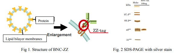
Basic type of BNC (BNC-L) is composed of L-protein of hepatitis B virus surface antigen which is floating on the particle made from lipid bi-layer. The BNC itself has ability to deliver its content into cytoplasm. Among three regions of L-protein, BNC-ZZ expresses zz-tag amino acids sequence in Pre-S1 region, the outmost region. Because zz-tag binds Fc region of antibodies, BNC-ZZ can binds to antibodies without affecting the antigen recognition site.
Thus, BNC-ZZ can be used for, depending on the antibody used, cell-specific DNA delivery in vitro, detection of antigen-antibody complex by using pre-labeled BNC-ZZ, and other applications.
Source: Yeast(Saccharomyces cerevisiae)
Appearance: Lyophilized white powder
Structure: Nano size articles having a protein floating in lipid bilayer. The protein contains zz-tag which binds to FC region of antibodies. The mean particle size is 40 to 50nm as determined dynamic light scattering methods (20 nm as determined by electron microscopy).
Content: 100 µg (dissolving instruction : added 500µL of water to 100 µg of lyophilized powder, that makes antigen concentration of 0.2 mg/mL in HEPES (10 mM HEPES、100mM NaCl, pH 7.2 - 7.4) containing 1% sucrose.
Protein purity: over 95 % (see SDS-PAGE data)
Storage: -20°C (stable for over 6 years)
Application notes
1. Cell specific transfection for DNA and others
2. Detection probe for antigen-antibody complex. For example, as a replacement of the secondary antibody in various systems, such as western blotting and ELISA.
1. Structure
2.Antibody-specific cellular transfection by DNA.
Mehods: Plasmid DNA was first mixed with a cationic liposome to form a DNA-liposome complex. The DNA-liposome complex was then mixed with BNC-ZZ to make a DNA-liposome-BNC-ZZ complex. To the complex, anti-EGFR antibody (mouse monoclonal) was mixed to form the transfection complex. The complex was applied on Gli36 cells (a glial cell line, expressing Fig 1. Structure of BNC-ZZ Fig. 2 SDS-PAGE with silver stain For Research Use Only Enlargement Protein Lipid bilayer membranes ZZ-tag kDa 97.4 66.2 45 31 BNC-ZZ Make 100ng abundant EGFR on the cell surface), and GFP expression was observed 72 hrs after application.
Result: As seen in below photos, GFP expression was seen in cells applied the complex attached with EGFR antibody-attached complex, but not with no antibody.
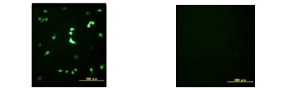
Fig. 3. Antibody-specific cellular transfection of DNA (GFP) into Gli36 cells using BNC-ZZ. Note: A similar antibody-specific transfection was succeeded in PC-12 with NGFR, MDA-MB453 with EGFR, A431 with FGFR and others. To achieve the antibody-specific transfection using BNC-ZZ, the choices of liposome and antibody species, and the composition ratio of BNC, liposome and antibody are critical. So to get better result, we strongly recommend taking screening processes to optimize these factors.
3.Simultaneous and multiple detection of proteins using fluorescent-labeled BNC-ZZ
Mehods: Four types of protein were separated by SDS-PAGE, and transferred to membrane from the gel. BNC-ZZ was separately labeled with 4 types of Cy dyes, and each Cy dye-labeled BNC-ZZ was incubated with one of the four antibodies that binds to the proteins. Thus, each fluorescent labeled BNC-ZZ forms a fluorescent probe that recognizes one of the four proteins. The protein-transferred membrane was simultaneously incubated with all the four types of probes in the same buffer. The Cy dye was detected using fluorescent detector at wavelengths corresponding to each Cy dye and graphically merged.
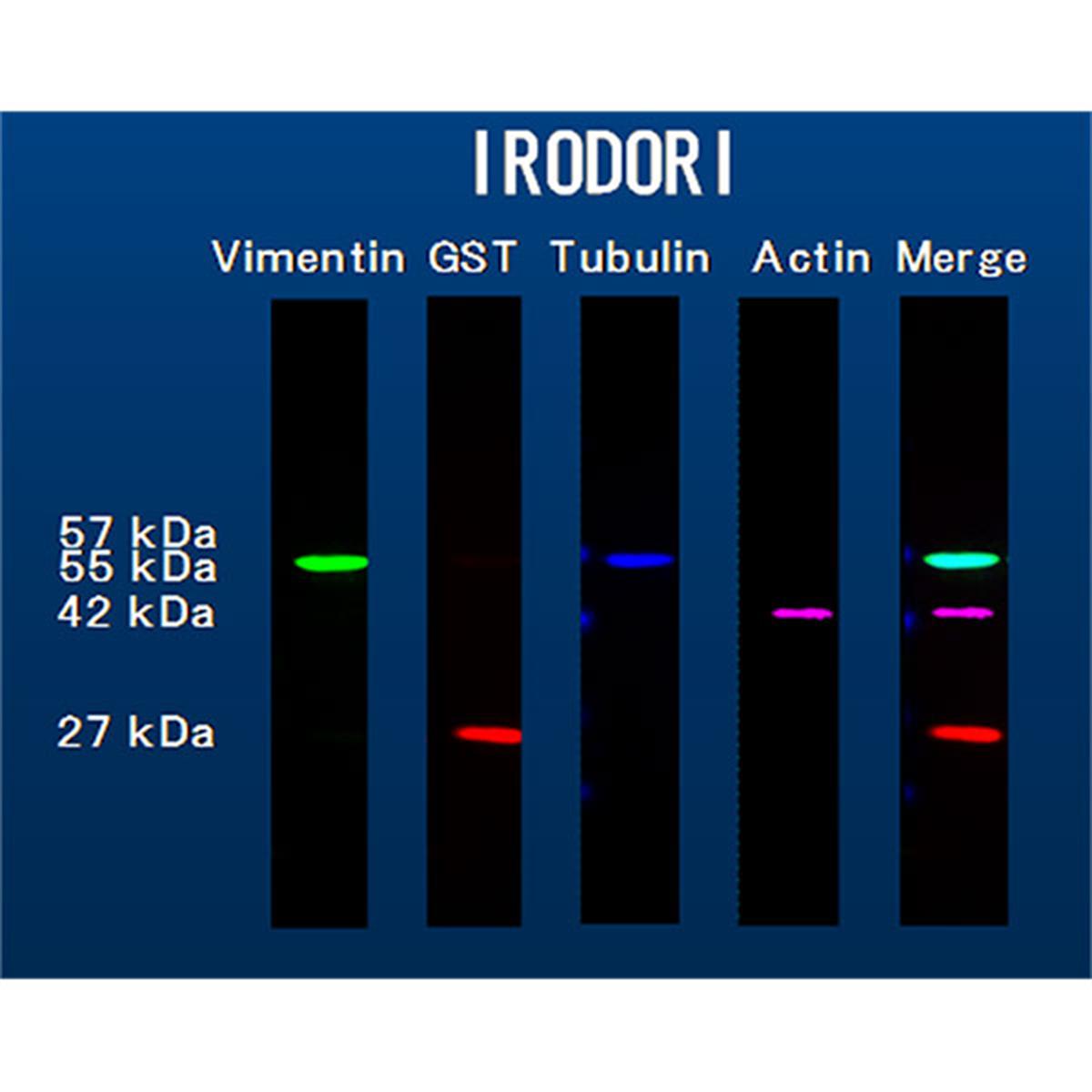
Result: As seen in below photos, four proteins (vimentin, GST, tublin, and actin) can be immuno-stained simultaneously without using second antibodies. The system has also an great advantages since same type of antibody, such as mouse IgG1, can be used for detecting multiple proteins.
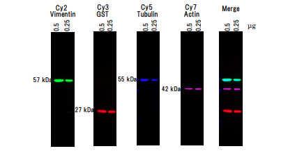
Fig. 4. Simultaneous and multiple color WB using BNC-ZZ Note: For performing this detecting system a kit is available on request. The kit is composed of BNC-ZZ (pre-labeled, or user labeld), blocking agents, and a detailed protocol.