


Anti-gamma tubulin(Xenopus)antibody, affinity-pure(rabbit), Azide & carrier free
Product#: FNK-69-001
$313.78
Availability:
Ships in 1-2 Weeks
Anti-gamma tubulin(Xenopus)antibody, affinity-pure(rabbit), Azide & carrier free
Cat.No: FNK-69-001Size: 100 μg
Storage: -20℃
Concentration: 1.0 mg/ml
Buffer: PBS- with 50% glycerol
Purity: Affinity-purified with immunogen.
Immunogen: Xenopus gamma-tubulin C-terminal peptide, C-EYHAATRPDYISWGTQDK conjugated with KLH
Isotype: Rabbit IgG
Reactivity: Xeno, human and rodents.
Application
1. Western blotting (200~1,000 fold dilution)2. Immunofluorescenct staining (1/100~1/200 dilution)
Background
Tubulin is the major constituent of microtubules. There are three members (α, β, γ) and two subtypes (δ, ε) in the tubulin family. Of these members, Gamma tubulin (451aa, 51KDa) is found at microtubule organizing centers (MTOC) such as the spindle poles or the centrosome, suggesting that it is involved in the minus-end nucleation of microtubule assembly during cell cycle.Data Link
UniProtKB/Swiss-Prot P23330 (TBG1_XENLA)Please note: All products are FOR RESEARCH USE ONLY. NOT FOR USE IN DIAGNOSTIC PROCEDURES. NOT FOR MILITARY USE.
Data Images
69-001 Anti-γ-tubulin antibody, rabbit polyclonal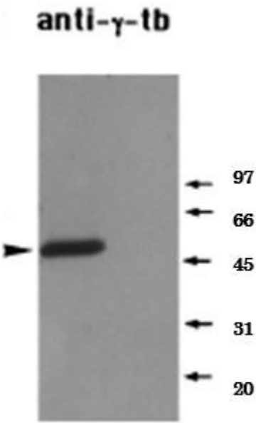
Fig. 1. Identification of endogenous γ-tubulin in a Xenopus mitotic extract by western blot
The primary antibody was used at 1/1,000 dilution.
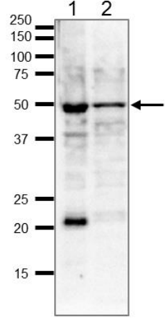
Fig.2 Identification of endogenous γ-tubulin in whole cell extracts by western blot
Samples ( 20 μg ) ; 1. HeLa cells, 2. MCF7 cells
Blotting was done with wet system overnight at 15 V.
The antibody was used at 1/1,000 dilution.
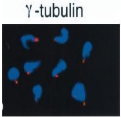
Fig.3 Immunofluorescent staining of γ-tubulin accumulated at sperm centriole in demembranated
Xenopus sperm heads which have been incubated in a Xenopus egg mitotic extract containing nocodazole. γ-Tubulin is stained red and sperm chromatin is stained with DAPI (blue). Anti-γ-tubulin (Xenopus) was used at 1/100 dilution.
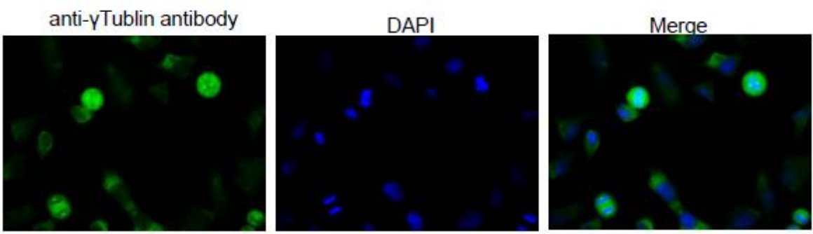
Fig.4 Immunofluorescence staining of γ-tubulin in Hela cells by using this anti-γ-tubulin antibody
The first antibody was used at 1/100 dilution. As second antibody goat anti-rabbit IgG conjugated with Alexa Fluor 488 was used at 1/1,000 dilution.
Reference
This product is described and used in the following reference.1. Masuda H et al Role of γ-tubulin in mitosis-specific microtubule nucleation from the Schizosaccharomyces pombe spindle pole body J. Cell Sci. 109,: 165-177(2000) PMID: 8834801 (WB)
2. Takeda S. et al. Identification of Ribonucleotide Reductase Protein R1 as an Activator of Microtubule Nucleation in Xenopus. Mol Biol Cell 11: 4173-4187 (2000) PMID: 11102516 (IF)