
Anti-Rad51(S.cerevisiae)Antibody, rabbit serum
Product#: FNK-62-101
$303.44
Availability:
Ships in 1-2 Weeks
Anti-Rad51(S.cerevisiae)Antibody, rabbit serum
Cat.No: FNK-62-101Size: 100 μg
Storage: -20℃
Concentration: 1.0 mg/ml
Buffer: PBS- with 50% glycerol
Purity: Affinity-purified with immunogen.
Immunogen: Purified recombinant His-tagged Sc Rad51 protein (full-size)
Reactivity: S. cerevisiae
Validation: S.cervisiae Rad 51 protein. The specificity of reaction was confirmed with rad51 mutant by WB (Fig.1)
Application
1.Western blotting (1/500~1/2,000 dilution)2. Immunoprecipitation
3. Chromatin Immuno-Precipitation (Assay dependent)
4. Immunofluorescence staining
5. ELISA
Background
S. cerevisiae Rad 51 protein (400 aa, 43 kDa) is a functional and structural homolog of E.coli RecA and human Rad51 proteins and plays a central role in DNA homologous recombination and recombination repair by promoting homologous DNA strand exchange reaction. Dmcl, Rad55, Rad57 are paralogs of Rad51 and they form complex with Rad51 and Rad52 in mediating recombination processes.Data Link
UniProtKB P25454 (RAD51_Saccharomyces cerevisiae)Please note: All products are FOR RESEARCH USE ONLY. NOT FOR USE IN DIAGNOSTIC PROCEDURES. NOT FOR MILITARY USE.
Data Images
62-101 Anti-Rad51 (S. cerevisiae) antibody, rabbit polyclonal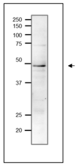
Fig.1 Western blot of endogenous Rad51 protein in crude extract of S, cerevisiae.
Proteins in the extract were separated on 12.5% SDS-PAGE and transferred to membrane in wet system overnight. The antibody was used at 1/1,000 dilution. As 2nd antibody, HRP conjugated goat anti-rabbit IgG antibody was used at 1/10,000 dilution.
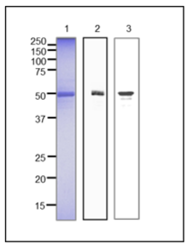
Fig.2 Western blot of recombinant scRad51 protein and yeast crude extract.
1. Recombinant Rad51 protein as analyzed by SDS-PAGE.
2. Western blot of recombinant scRad51 protein (10 ng).
3. Western blot of crude extract of S. cerevisiae strain BY4741.
For Western blot, anti-scRad51 antibody was used at 1/1,000 dilution.
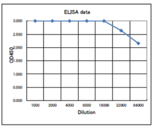
Fig.3 Titration of antibody reactivity of anti-Rad51 antibody by ELISA
Plate was coated with 100 μg of recombinant Rad51 protein (S. cervisiae) per well and 100 µl of the antiserum at the indicated dilution was added to each well and incubated. After washing, goat antirabbit-IgG conjugated with HRP was added as 2nd antibody. Color was developed with TMB as substrate.
References
This antibody was used in the following publications.1.Ribeyre C, Shore D. Anticheckpoint pathways at telomeres Nat Struct Mol Biol. 2012,2.19 : 307-13 PMID: 22343724 ChIP (S. cerevisiae)
2. Muramoto N et al. Phenotypic diversification by enhanced genome restructuring after induction of multiple DNA double-strand breaks. Nat Commun. 2018 May 18;9(1):1995.PMID:29777105. IF (S. cerevisiae)