Anti-PD-L1, Rat-Mono(6C11-3A11)
Product#: FNK-FDV-0054
$598.00
Availability:
Ships in 1-2 Weeks
Anti-PD-L1, Rat-Mono(6C11-3A11)
*Research use only, not for human or animal therapeutic or diagnostic use.Cat . No : FNK-FDV-0054
Description
- Size: 100 µL 0.5 mg/mL
- Formulation: Purified in PBS
- Host Species and Clonality: Rat monoclonal
- Immunogen: Bovine PD L1 extracellular region
- Clone name: 6C11 3A11
- Isotype and Subclass: IgG 2a,k
- Purification: Sequential ion exchange chromatography purified
- Lot Number: See vial label
- Specificity: Cow, Dog, Horse, Pig, Sheep, Goat, Buffalo
- Not reactive with Cat and Human
- Storage: For short-term storage, -20 °C For long-term storage, -80 °C storage is preferable.
- Avoid repeated freeze-thaw cycles and avoid storage at 4 °C.
Product Background
Programmed cell death 1 (PD-1) is a member of the immunoglobulin superfamily expressed mainly on T cells and inhibits T cell proliferation and effector functions. PD-L1, a ligand for PD-1, is known to be induced by various mechanisms, including inflammatory cytokines, and is highly expressed in many types of cancer cells. PD-L1 is attracting attention as an immune escape mechanism for cancer cells. Therefore, inhibition of PD-1 1/PD L1 interaction is expected to be a promising anti-tumor strategy, and basic research, drug discovery, and clinical studies are actively being conducted in humans and rodents, and several approved drugs have been launched. On the other hand, the function of PD-L1 in animals such as cattle and dogs has not been well analyzed. PD-L1 has high homology among animal species, suggesting its function may be conserved across species. The monoclonal antibody 6C11-3A11 against the bovine PD L1 extracellular region has been found to react with PD L1 in a wide range of animal species (see below “ Description ”). It is expected to be an excellent experimental tool for immunohistochemical staining and flow cytometry analysis. 6C11 3A11 is suitable for the detection and quantification of PD L1 in these animal species and is being used intensively to elucidate PD L1 function in each animal (ref. 1-11). For example, it has been used to observe infection and inflammation dependent PD L1 upregulation in cattle (ref. 1-4), to detect PD L1 expression in malignant tumors and to elucidate the mechanism of PD L1 expression induction by inflammatory cytokines in dogs (ref. 5-8), and to analyze stimulus dependent PD L1 expression responses in pigs (ref. 9), horses (ref. 10), and sheep (ref. 11).
Applications
-Immunohistochemistry (Paraffin)
-Flow cytometry
Recommended usage
| Immunohistochemistry | 1:100-1: 5 0 dilution |
| *Optimal dilutions should be determined by the researcher. | |
| Flow cytometry | 1:50 dilution |
| *Optimal dilutions should be determined by the researcher. |
Reference data
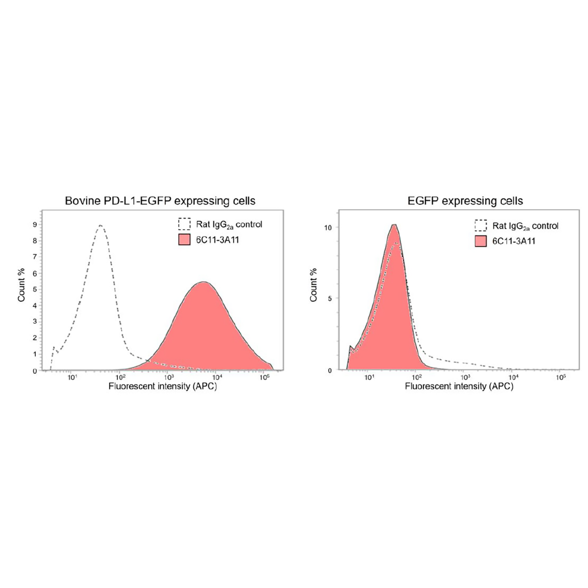
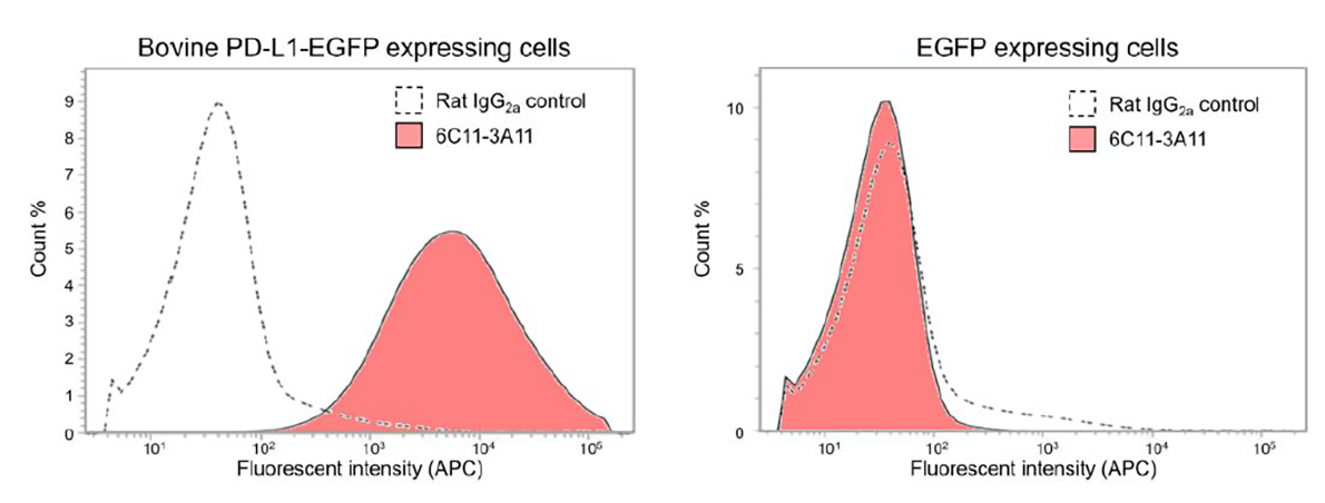
Validation for bovine PD L1
CHO cells overexpressing bovine PD-L1 fused with EGFP (PD L1 EGFP) or EGFP was treated with anti-PD-L1 antibody (6C11 3A11; 10 µg/ml) or rat IgG 2a isotype (10 µg/ml) as a negative control for 20 min at R.T. Then, cells were treated with anti-Rat Immunoglobulin ( allophycocyanin ( conjugated for 20 min at R.T. and applied to flow cytometry analysis. While 6C11 3A11 shows little binding to EGFP expressing cells, 6C11 3A11 significantly binds to bovine PD-L1 EGFP expressing cells.
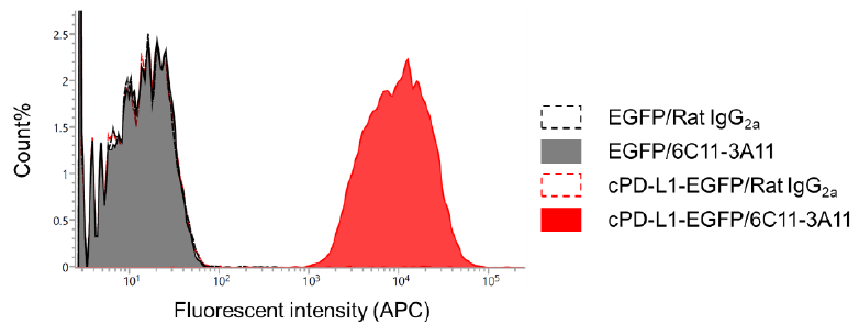
Validation for canine PD L1
CHO-DG44 cells overexpressing canine PD-L1 fused with EGFP (cPD L1 EGFP) or EGFP were treated with anti-PD-L1 antibody (6C11 3A11; 10 μg/ml) or rat IgG 2a isotype (10 µg/ml) as a negative control for 30 min at R.T. Then, cells were treated with anti-Rat Ig APC conjugated for 30 min at R.T. and applied to flow cytometry analysis. While 6C11 3A11 shows little binding to EGFP expressing cells, 6C11-3A11 significantly binds to canine PD L1 EGFP expressing cells.
Application examples
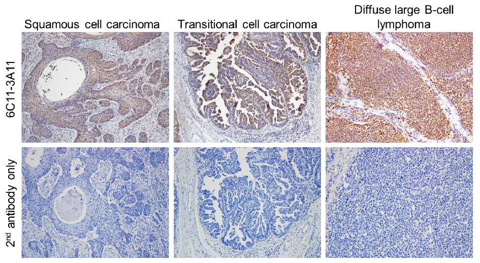
Immunohistochemistry of dog tumor tissues
Formalin-fixed and paraffin-embedded tissue sections of dog cancers, including squamous cell carcinoma, transitional cell carcinoma and diffuse large B-cell lymphoma, were sequentially treated with an anti-PD-L1 antibody (6C11 3A11 5 μg /ml and with polymer type rat IgG secondary antibody HRP conjugated and visualized by 3, 3-diaminobenzidine (DAB) staining. As a counterstain, Mayer’s hematoxylin stain was also performed. All tumor tissues show significant signals of 6C11-3A11 in cancer cells.
Flow cytometry analysis of cytokine stimulated dog cancer cells
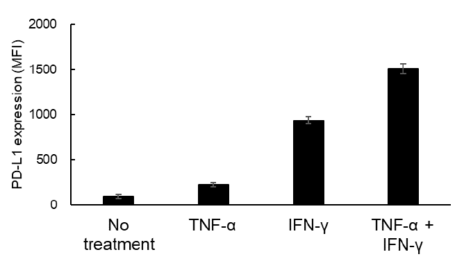
Canine malignant melanoma cell line CMeC (1.0 x 10⁵ cells/well) was stimulated with recombinant canine cytokines (TNF-α (10 ng/ml), IFN-γ (10 ng/ml) and TNF-α/ IFN-γ (10 ng/ml each) for 24 hours. The cells were incubated in 10% goat serum/PBS for 20 min R.T. as a blocking process and then stained with anti-PD-L1 antibody (6C11 3A11; 10 g/m l ) for 30 min R.T. After wash ing the cells with 10% goat serum/PBS, the cells were incubated with APC-conjugated anti-rat Ig secondary antibody for 30 min R.T. and finally washed twice. The labeled cells were analyzed by a flow cytometry system . The expression of PD-L1 was increased in TNF-α α or IFN-γ-treated cells, respectively, and the double treatment had a stronger effect on PD-L1 expression than the single treatment.
Reference
Cow
1.Sajiki et al., Infect Immun.Immun., 86 , e00910 17 (2018)
2.Sajiki et al., J. Immunol. Immunol., 203 , 1313 1324 (2019)
3.Goto et al., Front. Vet. Sci. Sci., 7 , 12 (2020)
4.Sajiki et al., Sci. Rep. Rep., 11 , 1063 (2021)
Dog
5.Takeuchi et al., Front. Vet. Sci. 7 , 330 (2020)
6.Maekawa et al., NPJ Precis. Oncol. Oncol., 5 , 10 (
7.Gulay et al., Sci. Rep. Rep., 12 , 2124 (2022)
8.Owaki et al., Vet.Comp. Oncol. Oncol., 21 , 279 290 (2023)
Pig
9.Ganbaatar et al., Immun. Inflamm. Dis. Dis., 9 , 1573 1583 (2021)
Equine
10.Ganbaatar et al., PLOS One 15 , e0234218 (2020)
Sheep
11.Tiyamanee et al., Vet. Immunol. Immunopathol . 261 , 110609 (2023)