

- Home
- Antibody
- Antigen I-K
- Anti-Influenza A Virus Nucleoprotein antibody, mouse monoclonal (C43), HRP-conjugated
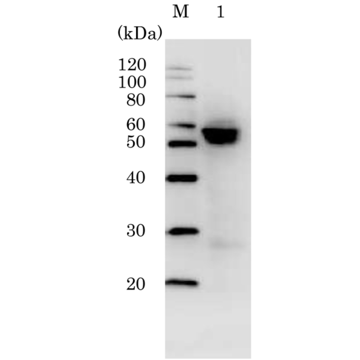
Anti-Influenza A Virus Nucleoprotein antibody, mouse monoclonal (C43), HRP-conjugated
Product#: FNK-65-111
$346.79
Availability:
Ships in 1-2 Weeks
Anti-Influenza A Virus Nucleoprotein antibody, mouse monoclonal (C43), HRP-conjugated
General Information
| Cat. No. | :FNK-65-111 |
| Size | :50 ug |
| Host Species | :Mouse |
| Label | :HRP |
| Purification | :Produced in serum-free medium and purified by proprietary chromatography procedure under mild conditions. 90~95% pure by SDS-PAGE |
| Reactivity | :Reacts with NP of all influenza A viruses tested, including seasonal H2N2, H3N2, and avian H5N1, H5N2 and H1N1 (seasonal, pandemic and swine). No cross reactivity with influenza B viruses. |
| Immunogen | :Human Influenza A Virus (H2N2) Okada strain |
| Isotype | :mouse IgG2A |
| Application |
|
| Storage | :Ship at 4℃, and store at -20℃. Do not freeze |
| Form | :1 mg/ml in PBS, 50% glycerol, filter sterilized |
Description
Influenza virus is an RNA virus, which causes influenza, and belongs to the family Orthomyxoviridae. Influenza virus is classified into three different genera, influenzavirus A, B, and C. They all have similar structures and compositions. The virions are 80-100nm in diameter and usually roughly spherical. The outer surface of the virion is made of a viral envelope containing two major glycoproteins, hemagglutinin (HA) and neuraminidase (NA). Influenzavirus A is further classified into subtypes based on the surface glycoproteins, HA and NA. Currently, there are 16 HA and 9 NA subtypes. The central core of the virion contains the viral RNA genome, which is packaged in the form of ribonucleoprotein complexes.
Influenza virus nucleoprotein (NP) is a major component of the ribonucleoprotein complex and is abundantly expressed during the course of infection. It is a structural protein, which encapsidates the negative strand viral RNA and is essential for RNA transcription, replication and packaging. NP binds the PB1 and PB2 subunits of the viral RNA polymerase and the matrix protein M1, in addition to its binding to ssRNA. NP is also known to interact with variety of other macromolecules of both viral and cellular origins, and these interactions have been shown to be essential for the viral lifecycle.

Fig.1. Western blotting of MDCK cells infected with H1N1 (A/PuertoRico/8/34), H5N1 (A/duck/HK/342/78), or H5N2 (A/crow/Kyoto/53/04) using C43 antibody.
Samples were collected at 3, 9, 24, and 48 hours post-infection. C43 detected NP after 3 hours post-infection and detected three different types of influenza viruses.
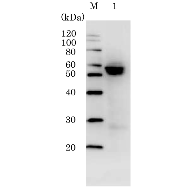 |
Fig.2 Western blotting of MDCK cells infected with H1N1 (A/PuertoRico/8/34) using HRP-conjugated C43 antibody. Proteins in the infected cell lysate was separated by 15% SDS-PAGE and blotted to PVDF membrane. The membrane was reacted with C43 monoclonal antibody conjugated with HRP at 1/1,000 dilution and visualized by Chemi-Luminescence. |
