


Anti-HIV-1 Protease, Rabbit-Poly
Product#: FNK-65-018
$303.44
Availability:
Ships in 1-2 Weeks
Anti-HIV-1 Protease, Rabbit-Poly
Cat.No: FNK-65-018Size: 100 μl
Storage: Store at 4℃ for short term. For long term storage store at -20℃. Aliquot to avoid repeated freezing and thawing.
Buffer: 0.09% Sodium azide
Purity: Rabbit antiserum
Immunogen: Full-size functional recombinant HIV-1 protease expressed and purified from E. coli
Isotype: Rabbit IgG
Reactivity: HIV-1 protease of all substrains
Application
1. Western blotting (1/2,000)2. Dot blotting (1/2,000)
3. Immunoprecipitation (1/2,000)
4. Inhibition of HIV-1 Protease activity (assay dependent)
5. ELISA (1/10,000)
Other applications have not been tested.
Background
HIV-1 protease is the aspartyl protease that mediates proteolytic cleavages of Gag and Gag-Pol polyproteins during or shortly after the release of the virion from the plasma membrane. Cleavages take place as an ordered, step-wise cascade to yield mature proteins. This process is called maturation. Displays maximal activity during the budding process just prior to particle release from the cell. Also cleaves Nef and Vif, probably concomitantly with viral structural proteins on maturation of virus particles. Hydrolyzes host EIF4GI and PABP1 in order to shut off the capped cellular mRNA translation. The resulting inhibition of cellular protein synthesis serves to ensure maximal viral gene expression and to evade host immune response.Data Link
UniProtKB P03367 (gag-pol), Q9YQ30 (HIV-1 Protease)Please note: All products are FOR RESEARCH USE ONLY. NOT FOR USE IN DIAGNOSTIC PROCEDURES. NOT FOR MILITARY USE.
Data Images
65-018 Anti-HIV-1 Protease antibody, rabbit serum 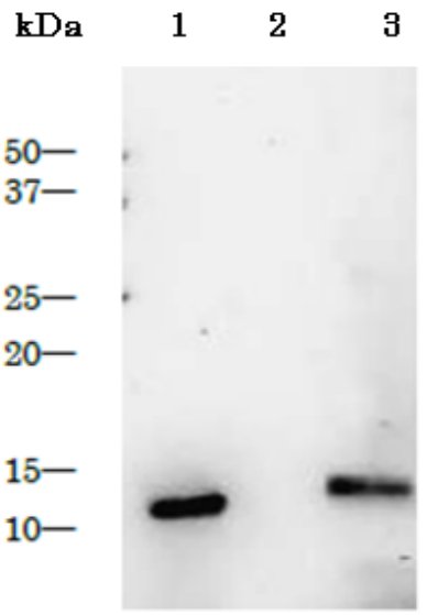
Fig.1 Detection of HIV-1 proteinase in the extract of HIV-1 infected cells by Western blotting using anti-HIV-1 protease antibody
1: Purified HIV-1 protease (1 ng, BioAcademia 05-013)
2: Extract of MT4 cells
3.: Extract of MT4 cells infected with HIV-1 (LAI strain)
The antiserum was used at 1/2,000 dilution.
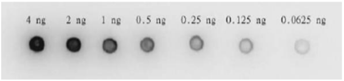
Fig.2. Dot blotting of HIV-1 protease by using anti-HIV-1 protease antibody
Anti-HIV-1 protease antibody was used at 1/2,000 dilution. As second antibody, goat anti-rabbit IgG antibody conjugated with HRP was used at 1/5,000 dilution. ECL system was used.
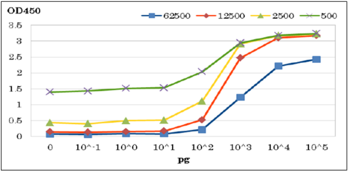
Fig.3 ELISA of HIV-1 protease with anti-HIV-1 protease antibody
The antibody was used at dilutions indicated above. Purified Protease was spotted on wells.
Reference
The HIV-1 strain and immunogen has been described in the following references.1. Adachi A et al “Production of acquired immunodeficiency syndrome-associated retrovirus in human and nonhuman cells transfected with an infectious molecular clone” J Virol 59: 284 - 291(1986) PMID: 3016298
2. Saitoh A et al “Overproduction of human immunodeficiency virus type I reverse transcriptase in Escherichia coli and purification of the enzyme” Microbiol Immunol 34:509-521 (1990) PMID: 1699113