Anti-GRWD1 antibody, rabbit polyclonal, ChIP grade
Product#: FNK-70-130
$346.79
Availability:
Ships in 1-2 Weeks
Anti-GRWD1 antibody, rabbit polyclonal, ChIP grade
General information
| Cat. No. | :FNK-70-130 |
| Size | :100 µg |
| Host Species | :Rabbit |
| Immunogen | :Purified GST-GRWD1 (human, full-length) expressed in E. coli. |
| Validation | :Validated for Western Blotting by siRNA. |
| KEYWORDS | :nucleolar stress response, oncogene, p53, PICT1, RPL11, tumor suppressor |
| Form | :1 mg/ml in PBS and 50% glycerol. Filter-sterilized. Carrier protein and azide free. |
| Reactivity | :Human, mouse and rat. Other species have not been tested. |
| Purification | :The antiserum was first absorbed with GST-agarose column and then the pass-through fraction was affinity-purified with GST-GRWD1 agarose column. |
| Application | :1. Western blotting (1/1,000~1/3,000 dilution) :2. Immunoprecipitation (Assay dependent) :3. Chromatin Immuno-Precipitation (Assay dependent) :4. Immunofluorescence staining / Immunochemistry (1/100~1/1,000 dilution) |
| Storage | :Ship at 4℃and store at -20℃. Do not freeze |
| Data Link | :uniprot/Q9BQ67 (GRWD1_HUMAN), uniprot/Q810D6 (GRWD1_MOUSE) |
Description
Glutamate-rich WD repeat-containing protein 1 (GRWD1) consists of 446 amino acids with molecular mass of 49.4 kDa. It has been found as a protein which interacts with METTL18 and CDT1 proteins. It has been implicated in regulation of DNA replication and/or in ribosome biogenesis.
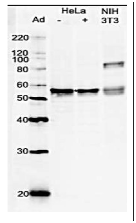 |
Fig.1. Identication of GRWD1 proteins in whole cell lysates by western blotting with anti-GRWD1 antibody
Whole cell lysates of HeLa cells untreated (-) and treated (+) with DNA damaging agent, adriamycin (Ad), and NIH3T3 cells were analyzed by western blotting with anti-GRWD1 antibody at 1/1,000 dilution. The samples were 10 μg. Second antibody was HRP-conjugated goat anti-rabbit IgG used at 1/5,000 dilution. The revelation of multiple bands indicates post-translational modification such as phosphorylation. The level of GRWD1 in the cell was not affected by DNA-damaging treatment. The identity of an additional band at ~85 kDa position other than the GRWD1 band in NIH-3T3 cell lysate is not known. The GRWD1 proteins were identified at a position higher (~55 kDa) than expected from the molecular mass of GRWD1 indicated from cDNA sequence (49.4 kDa). |
 |
|
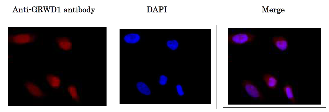
| Fig.2. Immunofluorescence staining of GRWD1 protein in HeLa cells. Hela cells were fixed in 4% paraformaldehyde overnight and permeabilized in 0.25% TritonX 100 in PBS for 10 min. Anti-GRWD1 antibody was used at 1/1,000 dilution. As second antibody, goat anti-rabbit IgG conjugated with Alex488 was used at 1/5,000 dilution. As a signal enhancer, Can Get Signal Immunostain B (Toyobo, Osaka) was used according to the protocol of the supplier. Nuclei were stained with DAPI. GRWD1 protein is localized in nuclei. |
|
 |
|
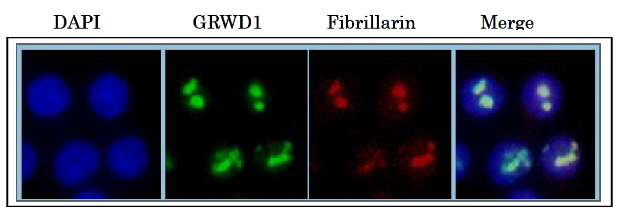
| Fig.3. Nucleolar localization of GRWD1. HCT116 cells were first extracted with Triton X-100 to remove nucleoplasmic proteins, double-immunostained with anti-GRWD1 (green) and anti-fibrillarin (red) antibodies as a marker for nucleoli, and counterstained with DAPI. The anti-GRWD antibody was used at 1/500 dilution and as the second antibody, goat anti-rabbit IgG conjugated with Alex488 was used at 1/2,000 dilution. |
|
References
This product has been described in Ref.1 and used in the following publication.
This product has been described in Ref.1 and used in the following publication.
- Sugimoto N. et al. Identification of novel human Cdt1-binding proteins by a proteomics approach: proteolytic regulation by APC/CCdh1. Mol Biol Cell. 2008 19(3):1007-21. WB
- Sugimoto N. et al. Cdt1-binding protein GRWD1 is a novel histone-binding protein that facilitates MCM loading through its influence on chromatin architecture. Nucleic Acids Res. 2015 Jul 13;43(12):5898-911. PMID:25990725 WB, IP, ChIP, IC/IF (human)
- Aizawa M. et al. Nucleosome assembly and disassembly activity of GRWD1, a novel Cdt1-binding protein that promotes pre-replication complex formation. Biochim Biophys Acta. 2016 Nov;1863(11):2739-2748. PMID:27552915 WB (human)
- Kayama K. et al. GRWD1 negatively regulates p53 via the RPL11-MDM2 pathway and promotes tumorigenesis. EMBO Rep. 2017 Jan;18(1):123-137. PMID:27856536 WB,IF, IP (human)
Aliases for GRWD1 Gene
- Glutamate Rich WD Repeat Containing 1 2 3 5
- WDR28 2 3 4
- GRWD 2 3 4
- Glutamate-Rich WD Repeat-Containing Protein 1 3 4
- RRB1 2 3
- Regulator Of Ribosome Biogenesis 1 Homolog 3
- CUL4- And DDB1-Associated WDR Protein 4 3
- Glutamate Rich WD Repeat Protein GRWD 3
- WD Repeat Domain 28 3
- KIAA1942 4
- GRWD1 5
- CDW4 3