

Anti-ATF6α, Mouse-Mono(1-7)
Product#: FNK-73-500
$315.24
Availability:
Ships in 1-2 Weeks
Anti-ATF6α, Mouse-Mono (1-7)
General information
| Cat. No. | :FNK-73-500 |
| Size | :50 ug |
| Host Species | :Mouse |
| Label | :Unlabeled |
| Specificity | :Specific to human ATF6α, no cross reactivity with mouse ATF6α |
| Immunogen | :Recombinant ATF6α (His-tagged amino-terminal fragment of ATF6α) |
| Isotype | :Mouse IgG2a k |
| Application | :Western blotting, Immunoprecipitation (IP), This antibody does not work for immunofluorescence analyses. |
| Storage | :Shipped at 4℃ or -20℃ and stored at -20℃. |
| Form | :Purified monoclonal antibody (IgG) 1mg/ml in PBS, 50% glycerol, filter-sterilized |
| Data Link | :UniProtKB/Swiss-Prot P18850 (ATF6A_HUMAN) |
Description
ATF6 (activating transcription factor 6) is an endoplasmic reticulum (ER) membrane-bound transcription factor activated in response to ER stress. When unfolded proteins accumulate in the ER, ATF6 is cleaved by regulated intramembrane proteolysis. The resulting amino-terminal fragment translocates to the nucleus and activates transcription by binding to ER stress-response elements present in the promoter regions of ER stress-inducible genes including those encoding ER chaperones and components of ER-associated degradation. The mammalian ATF6 family consists of two closely related homologs, ATF6a and ATF6ß. ATF6a but not ATF6ß plays a pivotal role in transcriptional control.
The monoclonal antibody was characterized in the laboratory of Professor Kazutoshi Mori of Kyoto University. The antibody was produced from hybridoma cultured in serum-free medium and purified under mild conditions by propriety chromatography processes.
Protocol for ATF6α analysis using anti-human ATF6α monoclonal antibody
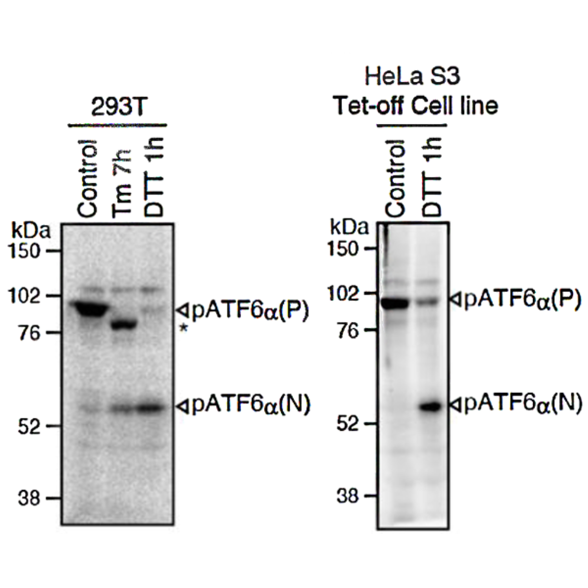
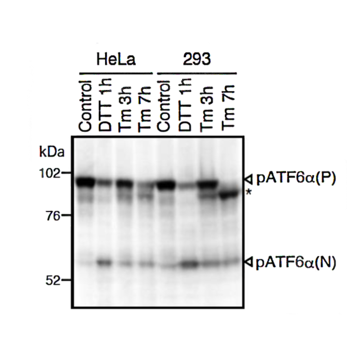
Both endogenous precursor ATF6α, pATF6α(P), and its cleaved product, pATF6α(N), can be detected in human cells such as HEK293T, HEK293 and HeLa cells by western blot analysis using anti-human ATF6α monoclonal antibody clone 1-7 (Fig. 1), according to the procedures described below. Clarity of the results may depend on cell types and culture conditions. If clear results could not be obtained by western blot analysis as seen with HeLa and HEK293 cells (Fig.1 right panel), it is worth while trying immunoprecipitation followed by western blot analysis according to the procedures described below.
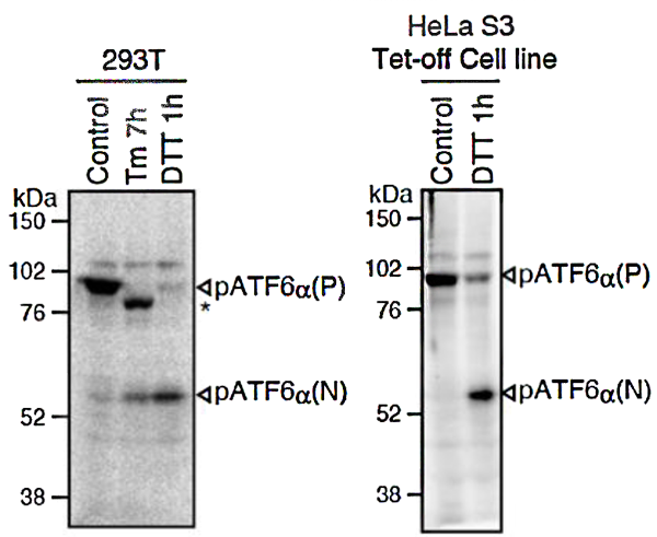
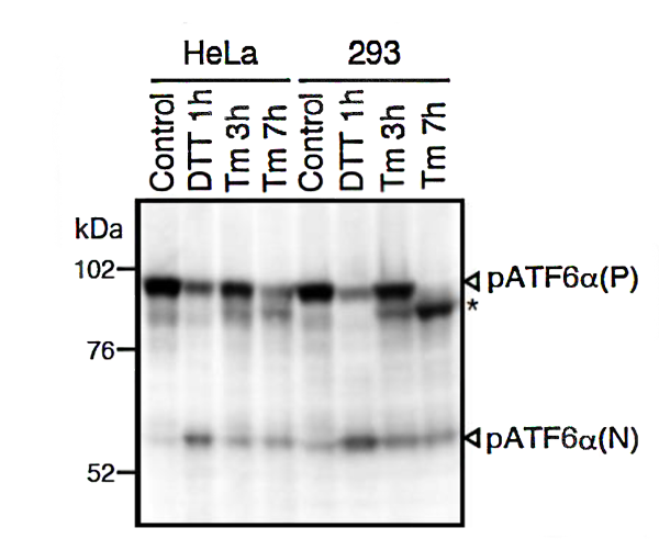
Fig.1 Western blot analysis of human cell extracts using this antibody: Conversion of pATF6α(P) to pATF6α(N) in DTT- or tunicamycin-treated cells.
Tm: 2 µg/ml tunicamycin (inhibitor of N-glycosylation). DTT: 1mM dithiothreitol (reducing reagent). The asterisk denotes an unglycosylated form of pATF6α (P).
ATF6α is constitutively expressed as pATF6α(P) (~90-kDa protein), and converted to pATF6α(N) (>50-kDa protein) in ER-stressed cells.
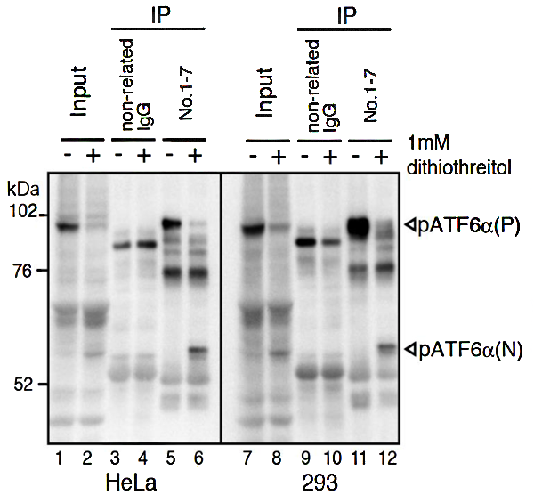
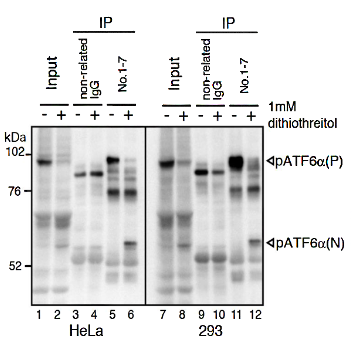
Fig.2 IP-Western blot analysis of human cell extracts using this antibody.
ATF6α was detected by Western blot (Input; lanes 1, 2, 7, and 8) using this antibody (No.1-7). After immunoprecipitation (IP) with non-related IgG (IP; lanes 3, 4, 9, and 10) or this antibody (No. 1-7) (IP; lanes 5, 6, 11, and 12), samples were subjected to SDS-PAGE and analyzed by Western blot using this antibody (# 1-7) and anti-mouse IgG antibody (light chain specific).
Detection of pATF6α(P) and pATF6α???????(N) is better in IP-Western blotting than Western blotting.
Antigen Characterization
| Type | :Transmembrane protein |
| Application | :Western blotting, Immunoprecipitation, Immunofluorescence |
| Species | :Human |
| Recombinant | :Yes |
ATF6α in the Cell
- Membrane-anchored transcription factor located in the endoplasmic reticulum (ER).
- Manages the unfolded protein response (UPR) by addressing misfolded protein accumulation in the ER.
- Transports to the Golgi apparatus for proteolytic cleavage when misfolded proteins accumulate.
- The N-terminal fragment translocates to the nucleus, activating genes that enhance protein folding capacity.
- Upregulates ER chaperones, foldases, and proteins involved in calcium regulation and redox balance.
- Influences cell survival by adapting protein folding capacity during ER stress.
- Promotes cell survival and proliferation, particularly in secretory cells like pancreatic beta cells and neurons.
- Involved in cell cycle regulation and can drive cancer cell proliferation under certain conditions.
ATF6α in the Human Body
- Integral to maintaining tissue homeostasis and responding to physiological stress.
- Essential for normal organ development and function; embryonic lethality observed in ATF6α/β double knockout models.
- Implicated in protective roles, such as cardiomyocyte survival in response to cardiac stress and neuroprotection in brain ischemia.
- Provides cytoprotection in glomerular diseases.
- Deregulated activity can contribute to pathological conditions, including hepatocarcinogenesis and survival of dormant cancer cells.
- Mitigates damage from myocardial ischemia/reperfusion injury and supports cardiac function under stress conditions.
- Extends beyond cellular protein folding to broader implications in tissue health and disease resilience.
Aliases for ATF6 Gene
- Activating Transcription Factor 6 2 3 5
- Cyclic AMP-Dependent Transcription Factor ATF-6 Alpha 3 4
- CAMP-Dependent Transcription Factor ATF-6 Alpha 3 4
- Activating Transcription Factor 6 Alpha 2 4
- ATF6A 2 3
- ATF6-Alpha 4
- ACHM7 3
- ATF6 5

References
- Hai T et al “Transcription factor ATF cDNA clones: an extensive family of leucine zipper proteins able to selectively form DNA-binding heterodimers” Genes Dev 3: 2083-2090 (1989) PMID 2516827
- Haze K et al "Mammalian transcription factor ATF6 is synthesized as a transmembrane protein and activated by proteolysis in response to endoplasmic reticulum stress" Mol Biol Cell 10: 3787-3799 (1999) PMID: 10564271
- Yamamoto K et al “Transcriptional induction of mammalian ER quality control proteins is mediated by single or combined action of ATF6? and XBP1” Dev Cell 13: 365-376 (2007) PMID: 17765680
- Maiuolo J et al “Selective activation of the transcription factor ATF6 mediates endoplasmic reticulum proliferation triggered by a membrane protein” PNAS 108: 7832-7 (2011) PMID: 21521793 5. Mori K “Divest yourself of a preconceived idea: transcription factor ATF6 is not a soluble protein!” Mol Biol Cell 21: 11435-8 (2010) PMID: 20219975
- Sharma, Rohit B et al. “Atf6α impacts cell number by influencing survival, death and proliferation.” Molecular metabolism vol. 27S,Suppl (2019): S69-S80. doi:10.1016/j.molmet.2019.06.005
- Correll, R.N., Grimes, K.M., Prasad, V. et al. Overlapping and differential functions of ATF6α versus ATF6β in the mouse heart. Sci Rep 9, 2059 (2019). https://doi.org/10.1038/s41598-019-39515-5
- Zhou H, Zhang T, Chen L, et al. The functional implication of ATF6α in castration-resistant prostate cancer cells. The FASEB Journal. 2023; 37:e22758. doi:10.1096/fj.202201347R
- 1, et al. “ATF6α Optimizes Long-Term Endoplasmic Reticulum Function to Protect Cells from Chronic Stress.” Developmental Cell, Cell Press, 4 Sept. 2007, www.sciencedirect.com/science/article/pii/S1534580707002663.