Amido Black 10B Staining Solution
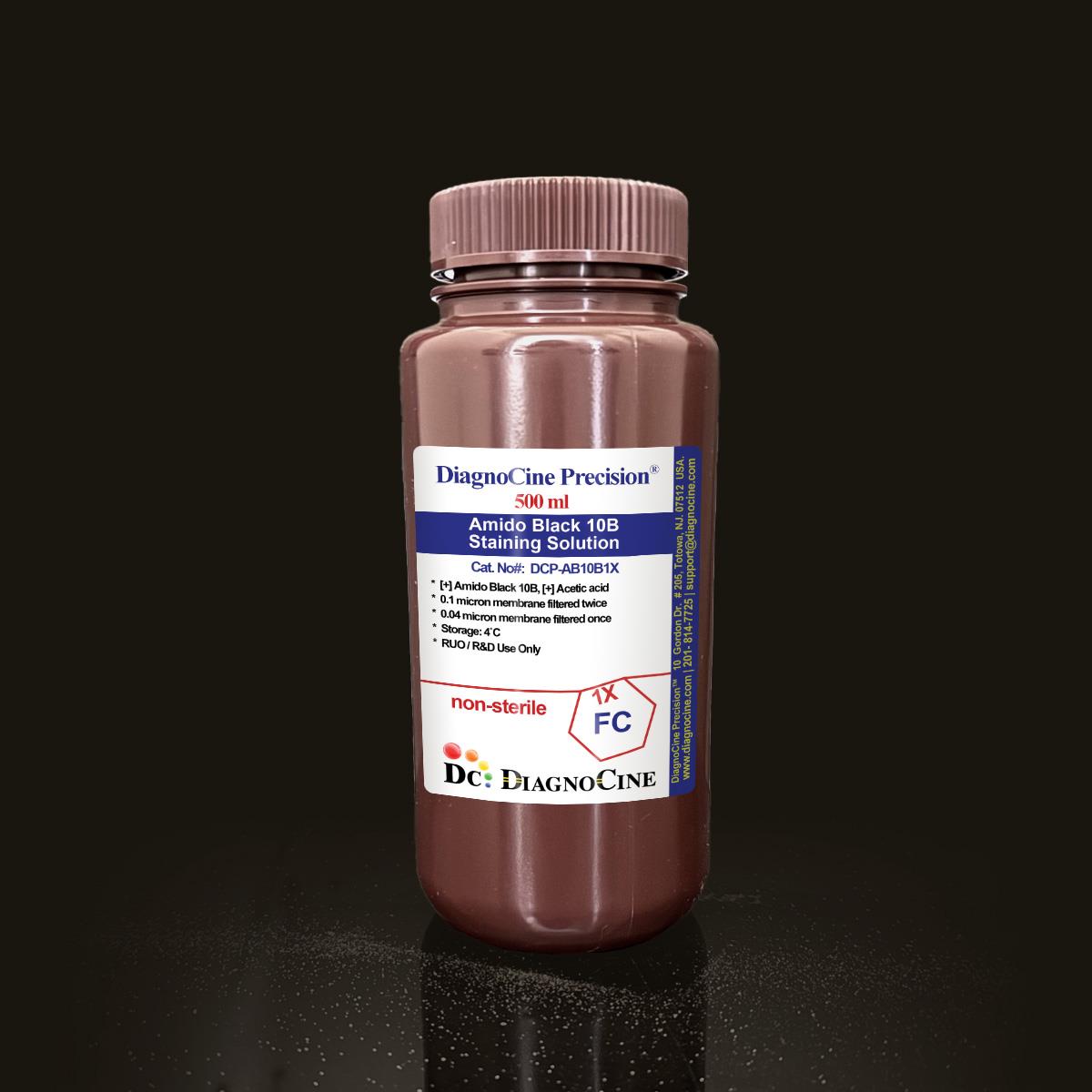
FluxMPS™ Amido Black 10B Staining Solution
A methanol-free, acetic acid-based (7%) formulation of Amido Black 10B (2%, CAS 1064-48-8) engineered for high-sensitivity total protein detection, forensic bloodstain enhancement, and collagen histology. Ultra-filtered through a 0.1 µm membrane twice and a 0.04 µm membrane twice for microchannel-safe, low-particulate dispensing across electrophoresis, blotting, and automated staining workflows.
- Filtered 0.1-micron membrane twice and 0.04-micron membrane twice — quadruple-stage purity architecture
- Methanol-free, acetic acid-based (7%) formulation eliminates flammable alcohol solvents
- Detects protein bands to 50 ng on nitrocellulose or PVDF membranes
- Blue-black staining reversible with 5–7% acetic acid destaining for downstream mass spectrometry or immunodetection
- DNase- and RNase-free per 18-hour incubation quality control
- Suitable for SDS-PAGE, Western blotting, forensic bloodstain enhancement, and collagen-specific histology
- Manufactured under ISO 13485-certified, CE-approved facilities
- Custom concentrations, pH, and additive formulations available on request
- pH2.5
- Formulation2% Amido Black 10B / 7% Acetic Acid
- AppearanceBlue-black solution
- Filtration0.1 µm x2 + 0.04 µm x2
- DNase ActivityNone detected (18 hours)
- RNase ActivityNone detected (18 hours)
- Storage4°C, protected from light
- Shelf Life6 months
- ManufacturingISO 13485-certified / CE-approved
- Intended UseResearch Use Only (RUO)
Engineered where standard staining reagents fall short
Conventional 0.22 µm-filtered stains and methanol-based formulations carry subvisible particulates, flammability risk, and background interference that compromise band resolution and bloodstain contrast. FluxMPS™ Amido Black 10B addresses each failure mode directly.
Microchannel-safe purity
Quadruple-stage 0.1 µm and 0.04 µm membrane filtration removes fine particulates that could otherwise interfere with electrophoretic band resolution or bloodstain pattern clarity.
Precise, stable pH
The acetic acid base (7%) has lower volatility than methanol, preventing concentration shifts over time and maintaining the formulation's pH stability throughout the labeled shelf life.
Ultrapure-grade water
Processed with Ultrapure Type 1 water (18.2 MΩ·cm) as part of the standard FluxMPS™ manufacturing process.
Low background for imaging & assays
The acidic composition ensures rapid dye penetration and minimal background interference, supporting high-resolution documentation of stained gels, blots, and bloodstain patterns.
Defined, traceable composition
A controlled 2% Amido Black 10B / 7% acetic acid formulation, with each lot subject to DNase and RNase quality control testing.
Customization on demand
Alternate concentrations, pH values, and additions of chemicals, compounds, proteins, or supplements are available upon request.
Quadruple-stage filtration system
FluxMPS™ Amido Black 10B Staining Solution is filtered through a 0.1-micron membrane twice and a 0.04-micron membrane twice, delivering a low-particulate reagent suitable for high-resolution electrophoresis, blotting, and forensic staining workflows.
-
1
0.1 µm Pre-filtration I
Removes large particulates and aggregates from the dye and acetic acid solution, extending downstream filter life.
-
2
0.04 µm Pre-filtration II
Retains fine particulates and bioburden below the size of the smallest known mycoplasma species, which measures about 0.2 micron.
-
3
0.1 µm Sterile-filtration I
A second 0.1 µm pass provides redundant particulate removal ahead of final polishing.
-
4
0.04 µm Sterile-filtration II — Final Polish
The second 0.04 µm pass delivers the final polish, helping prevent mycoplasma contamination of the staining solution.
Performance vs. conventional staining reagent
Sequential 0.1 µm and 0.04 µm membrane filtration, applied twice each, removes finer particulates than a single-pass filtration used in conventional staining reagents.

© Diagnocine® — DCP-AB10B1X
Where FluxMPS™ Amido Black 10B performs
From SDS-PAGE and Western blot total protein detection to forensic bloodstain enhancement and collagen-specific histology, this acetic acid-based formulation delivers consistent, low-background staining.
Automated Bioreactors & Robotics
An optional 0.01 µm (10 nm) ultra-filtered variant of this staining solution is available for laboratories running automated liquid handlers, gel stainers, and imaging robotics that require the lowest achievable particulate load.
- Total Particulate Exclusion for automated dispensing lines
- Valve & Sensor Protection in automated stainer/destainer systems
- Extended Stability during repeated automated staining cycles
Inquiry Required: the 0.01 µm (10 nm) ultra-filtered grade is available on request — contact support@diagnocine.com.
Micro Physiological System (MPS) & Chip
Low-particulate formulation suited to protein staining modules within microfluidic and lab-on-a-chip electrophoretic separation systems.
Wash, Dilution & Reconstitution
Working stain concentrations of 0.1–0.2% (w/v) Amido Black 10B in 7% acetic acid are readily prepared by dilution for SDS-PAGE and Western blot use.
SDS-PAGE & Agarose Gel Staining
Stains total protein in 5–15 minute protocols, with destaining completed in 2–24 hours — faster than the overnight destaining typically required with Coomassie Brilliant Blue.
Western Blotting & Downstream Detection
Fully reversible staining detects protein bands to 50 ng on nitrocellulose or PVDF membranes while remaining compatible with subsequent mass spectrometry or immunodetection workflows.
Latent Bloodstain Enhancement
Reacts with hemoglobin and serum albumin on porous and non-porous surfaces to produce permanent blue-black patterns, compatible with DNA preservation protocols.
Collagen-Specific Tissue Staining
Binds selectively to collagen fibers in tissue sections, enabling differential visualization of connective tissues in 10–15 minute protocols.
Specification summary
All figures below reflect this product's stated formulation and quality control data.
| Parameter | Specification |
|---|---|
| Formulation | 2% Amido Black 10B, 7% Acetic Acid |
| Appearance | Blue-black solution |
| pH | 2.5 |
| Parameter | Specification |
|---|---|
| Sterility Filtration | Filtered 0.1-micron membrane twice and 0.04-micron membrane twice |
| DNase Activity | None detected (plasmid DNA, 18 hours, room temperature) |
| RNase Activity | None detected (ribosomal RNA, 18 hours, room temperature) |
| Parameter | Specification |
|---|---|
| Storage Temperature | 4°C, away from bright light |
| Shelf Life | 6 months |
| Disposal | Neutralize with 1% sodium bicarbonate before disposal as hazardous waste |
| Parameter | Specification |
|---|---|
| Raw Materials Traceable | Amido Black 10B (CAS 1064-48-8), Acetic Acid |
| Manufacturing QMS | ISO 13485-certified and CE-approved facility |
| Traceability | Final packaging, QA, and testing at the Diagnocine R&D and Quality Testing Center; customization at Diagnocine Precision, Totowa, New Jersey, USA |
| Intended Use | Research Use Only (RUO) |
Full composition
A two-component acetic acid-based dye formulation, released against the concentrations below.
| Component | CAS Number | Concentration |
|---|---|---|
| Amido Black 10B | 1064-48-8 | 2% |
| Acetic Acid | 64-19-7 | 7% |
Manufacturing & compliance
Every lot of FluxMPS™ Amido Black 10B Staining Solution is produced, filtered, and released under a controlled quality system.
ISO 13485:2016 QMS
Manufactured under ISO 13485-certified and CE-approved facilities.
Ultrapure Type 1 Water
Processed with Ultrapure Type 1 water (18.2 MΩ·cm).
ISO Class 5 Fill & Finish
Final packaging and quality testing performed at the Diagnocine R&D and Quality Testing Center.
Micro-Batch Precision
Specific customization requests and assembly completed at Diagnocine Precision, Totowa, New Jersey, USA.
DNase Activity
None detected after 18-hour incubation with plasmid DNA at room temperature.
RNase Activity
None detected after 18-hour incubation with ribosomal RNA at room temperature.
Filtration Sterility
Filtered 0.1-micron membrane twice and 0.04-micron membrane twice.
Documentation / CoA
A Certificate of Analysis is available for this lot upon request.
How DCP-AB10B1X compares
A side-by-side look at the acetic acid-based FluxMPS™ formulation against a methanol-based Amido Black stain and against Coomassie Brilliant Blue.
| Parameter | DCP-AB10B1X (FluxMPS™) | Methanol-Based Amido Black | Coomassie Brilliant Blue |
|---|---|---|---|
| Methanol-Free Formulation | check_circle | cancel | check_circle |
| Number of Filtration Stages | 4 | Not specified | Not specified |
| Detection Sensitivity | 50 ng/band | Not specified | Not specified |
| Destain Time (Agarose Gel) | 2–24 hours | Not specified | Overnight |
| DNase/RNase-Free QC | check_circle | Not specified | Not specified |
| ISO 13485-Certified Manufacturing | check_circle | Not specified | Not specified |
| Custom Formulation Available | check_circle | Not specified | Not specified |
Frequently asked questions
Common questions about FluxMPS™ Amido Black 10B Staining Solution.
Supporting literature
Curated literature on Amido Black staining, protein blot detection, and forensic bloodstain enhancement.
- Wilson, C.M. Studies and critique of Amido Black 10B, Coomassie Blue R, and Fast Green FCF as stains for proteins after polyacrylamide gel electrophoresis. Analytical Biochemistry. doi:10.1016/0003-2697(79)90469-8
- Bickar, D.; Reid, P.D. A high-sensitivity protein assay based on Amido Black staining. Analytical Biochemistry. doi:10.1016/0003-2697(92)90276-4
- Sedmak, J.J.; Grossberg, S.E. A rapid, sensitive, and versatile assay for protein using Coomassie brilliant blue G250. Analytical Biochemistry. doi:10.1016/0003-2697(77)90370-2
- James, S.H.; Kish, P.E.; Sutton, T.P. Principles of Bloodstain Pattern Analysis: Theory and Practice. CRC Press. doi:10.1201/9781420005447
- Bergman, T.; Jornvall, H. Electroblotting of individual polypeptides from SDS/PAGE for direct sequence analysis. European Journal of Biochemistry. doi:10.1111/j.1432-1033.1987.tb13416.x
- Puchtler, H.; Waldrop, F.S.; Valentine, L.S. Polarization microscopic studies of connective tissue stained with picro-sirius red. Beitrage zur Pathologie. doi:10.1016/S0005-8165(73)80016-2
- Bhattacharyya, D.; Sarkar, S. Membrane filtration considerations for particulate control in laboratory reagent manufacturing. Journal of Membrane Science. doi:10.1016/j.memsci.2004.09.005
- Huang, Y.; et al. Microfluidic platforms for automated Western blot analysis. Lab on a Chip. doi:10.1039/C0LC00363H