

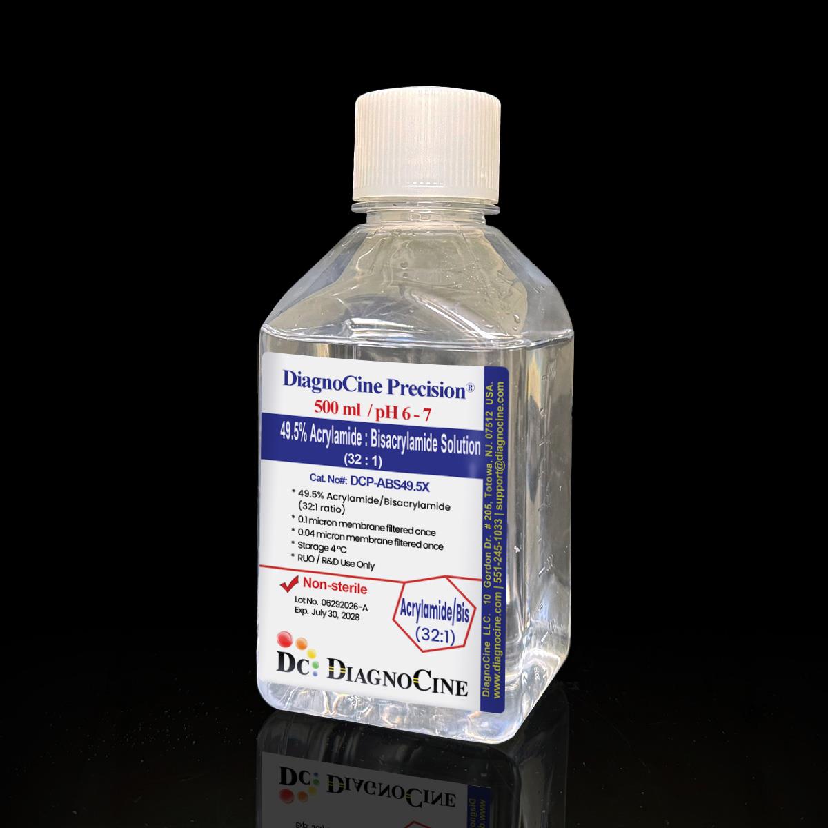
FluxMPS™ 49.5% Acrylamide : Bisacrylamide Solution (32:1)
Microfluidics Suitable, dual-stage ultra-filtered (0.1 µm x1 + 0.04 µm x1) 49.5% acrylamide:bisacrylamide (32:1) liquid solution engineered for high-resolution protein and nucleic acid electrophoresis. Ready-to-use, non-sterile grade formulation eliminates hazardous powdered-acrylamide handling while supporting on-chip and lab-on-chip gel-casting workflows.
- 49.5% total acrylamide (T) with 3% crosslinker (C) at a 32:1 acrylamide:bisacrylamide ratio, producing larger pore-size gels than standard 29:1 formulations
- Ready-to-use liquid solution eliminates the need to weigh and handle hazardous powdered acrylamide
- Dual-stage filtration: 0.1 µm membrane once, 0.04 µm membrane once, Non-Sterile grade
- No DNase activity detected after 18-hour incubation with plasmid DNA at room temperature
- No RNase activity detected after 18-hour incubation with ribosomal RNA at room temperature
- Clear solution, pH 6 - 7
- Manufactured under an ISO 13485-certified quality management system; final packaging, QA and customization performed at the Diagnocine R&D and Quality Testing Center, Totowa, New Jersey, USA
- Custom concentrations, tracking dyes, and pH modifications available on request
- Acrylamide:Bisacrylamide Ratio32:1
- Total Concentration (T)49.5%
- Crosslinker (C)3%
- pH6 - 7
- AppearanceClear solution
- Filtration0.1 µm x1 + 0.04 µm x1
- DNase ActivityNone detected
- RNase ActivityNone detected
- Storage4°C
- Shelf Life2 years from date of manufacture, unopened
Engineered where standard acrylamide solutions fall short
Conventional single-pass filtered acrylamide:bisacrylamide solutions carry particulate loads and inconsistent ratio control that show up as streaking, uneven pore structure, and batch-to-batch variability. FluxMPS™ addresses each failure mode directly.
Dual-stage filtered clarity
Sequential 0.1 µm and 0.04 µm filtration removes particulates and microaggregates that a single-pass 0.22 µm filter leaves behind, supporting clean gel casting.
Precision 32:1 formulation
Consistent 49.5% total monomer (T) and 3% crosslinker (C) ratio, batch after batch, so pore size and gel behavior remain predictable across experiments.
Nuclease-tested purity
No DNase or RNase activity detected after 18-hour incubation with plasmid DNA and ribosomal RNA, respectively, protecting nucleic acid samples run on these gels.
Low background for imaging
Clear, particulate-reduced solution supports clean band visualization during electrophoretic and imaging workflows.
Ready-to-use safety
Pre-mixed liquid formulation eliminates the need to weigh and handle hazardous powdered acrylamide in the lab.
Customization on demand
Alternate concentrations, tracking dye additions, and pH modifications are available — contact support@diagnocine.com.
Dual-stage filtration system
This solution is processed through a validated 0.1 µm prefilter followed by a 0.04 µm final filter, a Non-Sterile grade dual-stage train that removes particulates and microaggregates well below a conventional 0.22 µm single-pass filter.
-
1
0.1 µm Prefiltration
Removes large particulates and process residues, protecting the downstream 0.04 µm final filter cartridge.
-
2
0.04 µm Final filtration — Polish
Sub-micron polishing pass removes fine particulates and microaggregates, yielding a clear, ready-to-use solution suitable for high-resolution gel casting.
Filtration architecture
A dedicated prefilter protects the final filter cartridge in a two-stage, single-pair train — not a downstream cascade.
Where a 32:1 ratio solution earns its place
The larger pore size of a 32:1 acrylamide:bisacrylamide ratio suits separation of larger proteins and nucleic acid fragments, while the dual-stage filtration supports clean casting for microfluidic and lab-on-chip electrophoretic formats.
Automated Bioreactors & Robotics
For automated liquid-handling and robotic gel-casting systems, an optional 0.01 µm (10 nm) ultra nano-filtered MPS Grade variant of this solution is available on inquiry.
- Total Particulate Exclusion — reduces valve and nozzle fouling in automated dispensing systems
- Valve & Sensor Protection — minimizes particulate accumulation in microfluidic casting hardware
- Extended Perfusion Stability — supports longer unattended run cycles in automated platforms
Inquiry Required: the 0.01 µm MPS Grade variant is available by request — contact support@diagnocine.com.
SDS-PAGE & Native PAGE
Larger pore gels for improved resolution of high molecular weight protein species.
DNA/RNA Fragment Separation
Nuclease-tested formulation supports clean separation of nucleic acid fragments across a range of sizes.
On-Chip Gel Electrophoresis
Dual-stage filtered clarity suits gel casting within microfluidic and lab-on-chip electrophoretic devices.
Hydrogel Matrix Casting
Low-particulate solution supports reproducible hydrogel matrix casting for biosensor and microarray substrates.
High-Resolution Protein Separation
32:1 ratio offers a distinct pore profile useful for resolving large protein complexes and isoforms.
Point-of-Care Assay Development
Ready-to-use format suits gel-based assay development for point-of-care and lab-on-chip diagnostic platforms.
Full specification sheet
Every parameter below is stated exactly as verified for this product; parameters not established for this formulation (endotoxin, osmolality, CO2 requirement, sodium bicarbonate content) are intentionally omitted rather than estimated.
| Parameter | Specification |
|---|---|
| Formulation | 49.5% Acrylamide:Bisacrylamide, 32:1 ratio |
| Appearance | Clear solution |
| pH | 6 - 7 |
| Total Acrylamide (T) | 49.5% |
| Crosslinker (C) | 3% |
| Acrylamide:Bisacrylamide Ratio | 32:1 |
| Physical Form | Liquid |
| Parameter | Specification |
|---|---|
| Sterility | Non-Sterile; 0.1 µm x1 + 0.04 µm x1 filtered USP <71> |
| Mycoplasma | 0.1 µm mycoplasma-retentive filtration (not tested per lot) |
| DNase Activity | None detected (plasmid DNA, 18 hr, room temperature) |
| RNase Activity | None detected (ribosomal RNA, 18 hr, room temperature) |
| Manufacturing Standard | ISO 13485:2016 ISO 13485 |
| Parameter | Specification |
|---|---|
| Storage | 4°C |
| Shelf Life | 2 years from date of manufacture, unopened |
| Shipping Condition | Room temperature |
| Parameter | Specification |
|---|---|
| Traceability | Final packaging, QA and customization at Diagnocine Precision, Totowa, NJ, USA |
| Manufacturing QMS | ISO 13485-certified supplier facilities |
| UNSPSC | 41116155 — Molecular biology and cell culture growth media (UNv260801) |
| Regulatory Alignment | ISO 13485:2016 aligned |
| Production Method | Ready-to-use liquid formulation, dual-stage filtered |
| Intended Use | Research Use Only (RUO) |
Full composition
Total: 2 active components (Acrylamide, N,N'-Methylenebisacrylamide) formulated to a 32:1 ratio, T = 49.5%, C = 3%. Composition is provided on a per-lot release basis.
| Component | CAS Number | Concentration |
|---|---|---|
| Acrylamide | 79-06-1 | 48 g/100 mL (48% w/v) |
| N,N'-Methylenebisacrylamide (Bis-acrylamide) | 110-26-9 | 1.5 g/100 mL (1.5% w/v) |
| Component | CAS Number | Value |
|---|---|---|
| Total Monomer Concentration (T) | — | 49.5% |
| Crosslinker Concentration (C) | — | 3% |
| Acrylamide:Bisacrylamide Ratio | — | 32:1 |
| Component | CAS Number | Value |
|---|---|---|
| Solvent (Water) | 7732-18-5 | Balance to 100 mL |
| pH | — | 6 - 7 |
| Appearance | — | Clear solution |
Manufacturing & compliance
Every lot is processed, filtered and tested to a defined release specification before packaging.
ISO 13485 QMS
Manufactured under an ISO 13485-certified supplier quality management system.
Dual-Stage Filtration
0.1 µm prefilter followed by 0.04 µm final filter for consistent, low-particulate solution.
Nuclease-Tested
No DNase or RNase activity detected per lot after 18-hour incubation testing.
Finished in Totowa, NJ
Final packaging, quality assurance and customization performed at the Diagnocine R&D and Quality Testing Center.
DNase / RNase Testing
None detected after 18-hour incubation with plasmid DNA and ribosomal RNA, respectively, at room temperature.
Filtration Verification
Dual-stage 0.1 µm + 0.04 µm filtration verified per lot.
pH & Appearance
Confirmed clear solution, pH 6 - 7, prior to release.
Documentation / CoA
Certificate of Analysis available on request, detailing lot number, appearance, pH, filtration and nuclease testing results.
- Appearance, clarity and pH
- DNase and RNase activity
- Filtration verification (0.1 µm + 0.04 µm)
How DCP-ABS49.5X compares
Comparison figures are shown as specification against published specification; parameters without a sourced comparison figure are marked "Not specified" rather than omitted.
| Parameter | DCP-ABS49.5X (FluxMPS™) | Comparison |
|---|---|---|
| Grade | Microfluidics Suitable | Not specified |
| Acrylamide:Bisacrylamide Ratio | 32:1 (T=49.5%, C=3%) | Not specified |
| Final Filtration Pore Size | 0.04 µm | Not specified |
| Number of Filtration Stages | 2 (dual-stage) | Not specified |
| Mycoplasma Barrier Filtration | 0.1 µm mycoplasma-retentive filtration | Not specified |
| Endotoxin (release specification) | Not specified for this product | Corning classical liquid media — < 0.25 EU/mL Sigma-Aldrich DMEM complete medium — ≤ 2 EU/mL Gibco classical DMEM — Not specified (recorded per lot) |
| DNase / RNase Activity | None detected | Not specified |
| Manufacturing QMS | ISO 13485:2016 | Not specified |
| Custom Formulation | check_circle Available on request | Not specified |
Comparison figures from published supplier specifications, accessed 2026-09-02. Suppliers that publish no numeric endotoxin specification are shown as "Not specified".
Frequently asked questions
Common questions about DCP-ABS49.5X, 49.5% Acrylamide:Bisacrylamide Solution (32:1).
Supporting literature
Curated references on polyacrylamide gel electrophoresis, crosslinker ratio effects, and reagent purity considerations.
- Laemmli UK. Cleavage of structural proteins during the assembly of the head of bacteriophage T4. Nature. 1970;227:680-685. doi:10.1038/227680a0
- Righetti PG. Determination of the isoelectric point of proteins by capillary isoelectric focusing. J Chromatogr A. 1997;772:1-2. doi:10.1016/S0021-9673(97)00204-1
- Hjerten S. Molecular sieve electrophoresis in a discontinuous voltage gradient. Arch Biochem Biophys Suppl. 1962;1:147-151. doi:10.1016/0003-9861(62)90284-3
- Rueden CT, et al. ImageJ2: ImageJ for the next generation of scientific image data. BMC Bioinformatics. 2017;18:529. doi:10.1186/s12859-017-1934-z
- Sambrook J, Russell DW. Molecular Cloning: A Laboratory Manual, 3rd ed. Cold Spring Harbor Laboratory Press; 2001.
- Bio-Rad Laboratories. A guide to polyacrylamide gel electrophoresis and detection. Technical bulletin, 2016.
- Chrambach A, Rodbard D. Polyacrylamide gel electrophoresis. Science. 1971;172:440-451. doi:10.1126/science.172.3982.440
- Duong-Ly KC, Gabelli SB. Gel electrophoresis of proteins. Methods Enzymol. 2014;541:35-45. doi:10.1016/B978-0-12-420119-4.00003-2
- Tinsley RA, et al. On-chip electrophoretic separation for microfluidic diagnostic platforms. Lab Chip. 2015;15:2144-2153. doi:10.1039/C5LC00126A
- Whitesides GM. The origins and the future of microfluidics. Nature. 2006;442:368-373. doi:10.1038/nature05058